THE HIP JOINT
The stability of the hip joint owes much to the depth and extent of the acetabulum, which is considerably increased by a fibrocartilaginous rim; it embraces a large part of the femoral head (Figure 24-1Z√).
The head is additionally secured against luxation by two ligaments. One, the ligament of the femoral head, is short and stout but is not peculiar in any important way. The other, the accessory ligament, is unique to the horse (and donkey) among domestic species. It begins as a detachment from the prepubic tendon and reaches the joint by following a shallow groove on the ventral aspect of the pubis; this leads it to the acetabular notch through which it passes to insert on the head (see Figure 21-2Z5'). The two ligaments together restrict both the range and the versatility of movement permitted to the joint. The restrictions on rotation and abduction are most severe; in practice, movement is almost confined to flexion and extension in a sagittal plane, which is a much more limited repertory than the geometry of the articular surfaces suggests. The stability of the joint is partly dependent on the tension exerted by the weight of the abdominal viscera pulling on the prepubic tendon and thus on the accessory ligament (p. 547).Although the joint capsule is quite capacious, its deep location makes it relatively difficult to access. When it must be punctured, the needle is introduced between the two parts of the greater trochanter and is directed horizontally and craniomedially, at an angle of about 40° to the transverse plane.
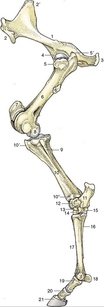
Figure 24-1 The skeleton of the left hindlimb, lateral view. 1, Hip bone (os coxae); 2, coxal tuber; 2', sacral tuber; 3, ischial tuber; 4, head of femur; 5, 5', cranial and caudal parts of greater trochanter; 6, femur; 6', third trochanter; 7, patella; 8, femoral condyle; 9, fibula; 10, tibia; 10', tibial tuberosity; 10", lateral malleolus; 11, calcaneus; 12, talus; 13, central tarsal; 14, third tarsal; 15, fourth tarsal; 16, metatarsal IV (lateral splint bone); 17, metatarsal III (cannon bone); 18, proximal sesamoid bones; 19, 20, 21, proximal, middle, and distal phalanges, the last within the hoof.