THE HOCK JOINT
The tarsocrural and proximal intertarsal joints, the only joint compartments at the hock accessible for injection, do not communicate. Two sites are available for injection of the tarsocrural joint, both on the lateral side: one is dorsal and the other is plantar to the collateral ligament.
The proximal intertarsal joint is entered from the medial side, plantar to the collateral ligament. There are two independent joint spaces at the tarsometatarsal level: one is proximal to metatarsals II and III, and the other is proximal to metatarsals IV and V. The first of these communicates with the distal intertarsal joint (Figure 36-4).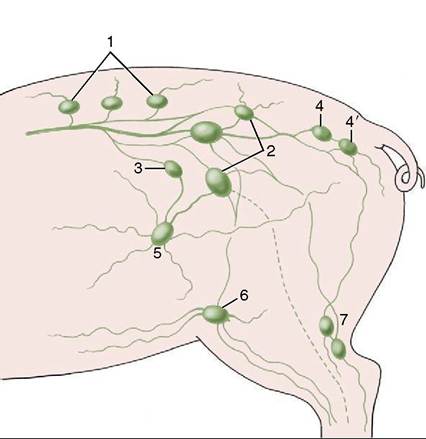
Figure 36-5 Lymph flow of the hindlimb, lateral view. 1, Lumbar aortic nodes; 2, medial iliac nodes; 3, lateral iliac node; 4, ischial node; 4, gluteal nodes; 5, subiliac nodes; 6, superficial inguinal nodes; 7, popliteal nodes.
CHAPTER 36

No account will be given of the arteries of the limb. Lymph from superficial structures of the thigh and leg drains to the superficial inguinal and subiliac nodes (Figure 36-5); that from deeper parts travels in lymphatic vessels that run with the major arteries to reach the medial iliac nodes. Lymph from the distal part of the limb drains to the popliteal nodes. Some efferents from these nodes proceed to the gluteal and ischial nodes on the lateral surface of the sacrosciatic ligament; others join the lymphatics running to the medial iliac nodes.