THE STIFLE JOINT
The three compartments of this joint communicate, which allows a single injection to reach all parts (see the dog in Figure 2-63 for the general idea). The puncture
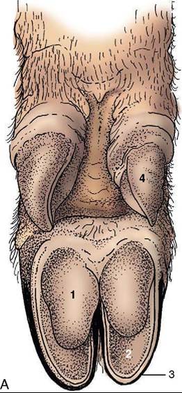
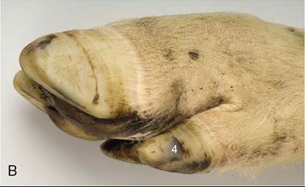
Figure 36-2 A, Palmar surface of the foot of a pig.
1, Bulb (digital pad) of hoof; 2, sole of hoof; 3, wall of hoof; 4, hoof of accessory digit. B, Lateral view of foot of a pig.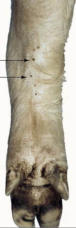
Figure 36-3 Carpal glands (arrows) of a pig, palmar view.
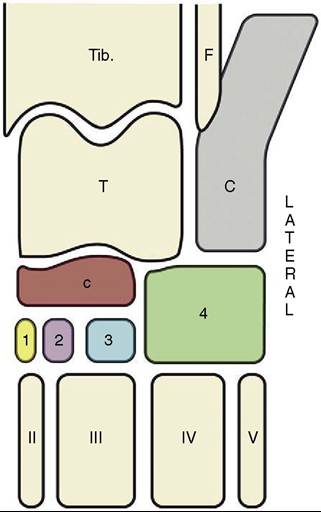
Figure 36-4 The bones of the tarsal skeleton in the pig, schematic. Roman numerals identify the metatarsal bones, Arabic numerals the distal tarsal bones. Tib, Tibia; F, fibula; T talus; C, calcaneus; c, central tarsal bone.
is made lateral to the patellar ligament, about one third of the distance down from the patella to the tibial tuberosity.
More on the topic THE STIFLE JOINT:
-
Veterinarian -