» The Hooves
The hooves of the principal digits curve toward each other at both ends, making contact behind and occasionally also at their apices (Fig. 30.10). The lateral hoof carries the greater share of weight and is larger than the medial one, although this is not always so in the hindfoot.
Each hoof consists of periople, wall, sole, and bulb. The ground surface is formed by the distal border of the wall, the sole, and the dorsal part of the bulb (Fig. 30.10/1, 3, and 4'); the parts visible in the standing animal are the wall to the sides and the bulb at the back of the hoof. The coronary border of the hoof is higher on the abaxial than on the axial side. The apical two thirds or so of the hoof are occupied by the distal phalanx and deep flexor tendon; the space behind is taken up by the digital cushion, the springy pad of fatty-fibrous tissue that also extends under the larger "half" of the bone (Fig. 30.11/8).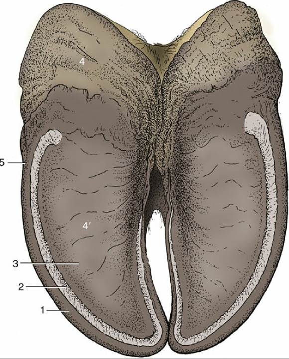
FIG. 30.10 Ground surface of the hooves of the bovine forefoot. 1, Wall; 2, white line; 3, sole; 4, bulb; 4', dorsal part of bulb; 5, abaxial groove on the wall, dividing wall from bulb.
Laminitis: The most important clinical disease of the claw in dairy cattle is subclinical laminitis. The signs include claw horn deformities, soft sole horn, and widened white line.
The periople provides a narrow (≈1 cm) strip along the coronary border that widens at the back where it grades into the bulb and merges with the periople of the other hoof. It is partly hidden by hair. In consistency it is intermediate between the epidermis of the skin and the hard horn of the wall.
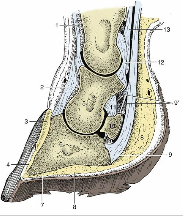
FIG. 30.11 Sagittal section of the medial digit of the bovine forefoot.
1, Proper (medial) digital extensor;2, common digital extensor; 3, coronary dermis; 4, laminar dermis; 5, middle phalanx; 6, distal phalanx; 7, sole dermis covered by sole; 8, digital cushion; 9, deep digital flexor; 9', fibers of deep digital flexor to the middle phalanx and navicular bone; 10, navicular bone; 11, collateral navicular ligament; 12, palmar ligaments of pastern joint; 13, superficial digital flexor.
The wall, sharply flexed on itself, forms the greater part of both axial and abaxial surfaces (Fig. 30.10); the flexure produces a crest at the front that curves distally toward the tip or "toe" of the hoof. Both surfaces are bounded caudally by more or less distinct grooves (Fig. 30.10/5) that extend from the coronary border to the ground surface; the horn caudal to the grooves belongs to the bulb. The axial groove is more cranial and provides an area of weakness that is sometimes penetrated leading to injection of the coffin joint located only a few millimeters away. The wall is marked by prominent ridges, parallel to the coronary border, caused by uneven production of horn due to local or more general disturbances. Although the distal border normally makes contact with, the ground along the whole length of the abaxial wall, it does so only toward the toe on the axial side; the greater part of this margin bears weight only on softer ground. The wall is thicker near the apex and toward the ground, especially abaxially. It consists of both tubular and intertubular horn and is produced over the wide, flat coronary dermis. The horny laminae are short and low and form a weaker union with the laminar dermis than in the horse. This may be correlated with the greater extent of the weight-bearing surface in ruminants.
The sole (Fig. 30.10/3) is a relatively smooth area confined within the inflected angle of the wall from which it is separated by the softer so-called white line. This line, hardly lighter than the unpigmented horn to each side, is only a few millimeters wide and comprises the alternation of the distal ends of the horny laminae with the slightly darker horn produced over the terminal papillae of the sensitive laminae.
Centrally, the sole blends imperceptibly with the apex of the bulb. The junction marks the extent of the digital cushion (Fig. 30.11/7 and 8).Sole Hemorrhage: Sole dermis is richly vascularized, and sole hemorrhages are a high-incidence clinical manifestation. The hemorrhages are associated with Iaminitis and rapid weight gain.
The bulb provides both the caudal aspect and a considerable portion of the ground surface where its apex inserts into the V-shaped sole. It is the chief weight-bearing part. A large proportion of intertubular horn makes it relatively soft, but its considerable thickness may compensate. Bulbar horn tends to flake when allowed to build up (as in animals that have stood on fouled bedding), and the resulting fissures provide access to infection leading to abscesses that may destroy the dermis and deeper structures.
The hoof capsule is molded on a dermis attached to underlying structures by a modified subcutis, best developed where it forms the digital cushion. The dermis presents segments that correspond to the parts of the hoof (Fig. 30.12). The horn of the wall is produced over the coronary dermis (Fig. 30.12/2) and slides distally over and between the dermal laminae, where horn just sufficient to maintain adhesion is produced.
The horn of other parts of the hoof grows away from the dermis at a rate of about 5 mm per month, with growth occurring a little faster in calves. In cattle allowed free range, wear at the ground surface equals growth, and at the toe the angle with the ground is maintained at about 50 degrees. On soft surfaces, growth exceeds wear, and the hooves must be trimmed periodically if the toe is not to grow forward at a lesser angle. When this occurs, the coffin joint is gradually overextended, the deep flexor tensed, and greater weight placed on the (caudal) part of the hoof over the insertion of the deep flexor and navicular bone. This causes pain and therefore lameness.
In late fetal life the distal parts of the hoof are covered with soft horn, which is said to prevent injury to the fetal membranes and the birth canal. This soft cushion soon dries when exposed to air.
The dewclaws, miniatures of the principal hooves, consist mainly of wall and bulb; they have no practical importance.