The Tendons
The interosseous muscle, morphologically a compound formation, is conventionally referred to in the singular (Fig. 30.7). This flat muscle is fleshy in the young but becomes increasingly fibrous as the animal matures and gains weight.
In the adult it forms a strong, almost wholly tendinous band that continues distally from the capsule of the carpal joint (Fig. 30.8/8). In midmetacarpus it gives rise to five principal branches; four of these—all but the central one—appear to terminate on the proximal sesamoid bones but obtain a functional continuation from the distal (sesamoidean) ligaments that attach on the proximal phalanges. The arrangement forms a "sling" that is tensed when the foot bears weight and the fetlock joint is overextended. Thin slips from the interosseous join the extensor tendons. Two of these split from the abaxial branches already mentioned and wind around the abaxial surfaces of the proximal phalanges to merge with the proper extensor tendons. Two more are provided by the bifurcation of the fifth (central) branch. They pass through the interdigital space, wind around the axial surfaces of the phalanges, and merge in the same tendons. In midmetacarpus the interosseous muscle also releases from its palmar surface a strong band (Fig. 30.8A/7) that divides to join the branches of the superficial digital flexor tendon above the fetlock. (The band may be regarded as a check ligament of the superficial digital flexor.)The three extensor tendons can be palpated where they lie side by side on the dorsal surface of the metacarpal bone. The middle tendon (from the lateral belly of the common digital extensor) bifurcates at the fetlock and the thin branches, each surrounded by an independent synovial sheath (Fig. 30.9/2'), follow the dorsal surface of the digits to insert on the extensor processes of the distal phalanges.
The medial tendon (from the medial belly) widens as it passes over the dorsal pouch of the fetlock joint, where a subtendinous bursa facilitates its passage. This tendon receives the extensor branches from the interosseous muscle before it inserts on the proximal end of the middle phalanx (but with a secondary connection to the distal phalanx). The lateral tendon (lateral digital extensor; Fig. 30.9/3) comports itself identically in relation to the lateral digit.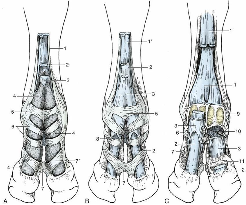
FIG. 30.7 Palmar view of the bovine forefoot. (A) Superficial dissection. (B) Tissues of the digital sheath have been removed. (C) Parts of the superficial and deep flexors have been removed. 1, Interosseous; 1', band of interosseous to superficial flexor; 2, deep digital flexor; 3, superficial digital flexor; 4, digital sheath;
5, annular ligament of fetlock joint; 6, digital annular ligaments; 7 and 7', distal interdigital ligament: 7, deep part, and 7', superficial part; 8, proximal interdigital ligament; 9, proximal sesamoid bones; 10, cruciate sesamoidean and interdigital phalangosesamoidean ligaments; 11, navicular bone.
The superficial and deep flexor tendons are separated from the metacarpal bone by the interosseous muscle (Fig. 30.8). Together they can be palpated as they emerge from the carpus medial to the accessory carpal bone, and they become individually distinguishable in the distal half of the cannon, where the deep fascia is thin. They are never so easily identified as the sharp-edged interosseous lying against the bone. The tendons are difficult to palpate in the digits.
The superficial flexor tendon splits above the fetlock joints (Fig. 30.7/3). Each branch receives a band from the interosseous muscle with which it forms a sleeve about the corresponding branch of the deep flexor when level with the proximal sesamoid bones. These bones provide bearing surfaces around which the combined tendons bend, secured in place by annular ligaments (Fig.
30.7/5 and 9). The palmar wall of the sleeve ends at the middle of the proximal phalanx, exposing the deep tendon that has now exchanged relative position with the superficial flexor. The dorsal wall of the sleeve continues the superficial flexor tendon and terminates on the proximal end and complementary cartilage of the middle phalanx. Two narrower (digital) annular ligaments strap the tendons to the proximal phalanx. The deep flexor tendon widens after leaving the confines of the sleeve and continues over the insertion of the superficial flexor tendon, which provides it with another bearing surface. The deep digital flexor tendon is protected by the navicular bursa during its passage over the navicular bone. It ends in a wide insertion on the hind end of the distal phalanx. The distal interdigital ligament binds the deep tendon down at the middle phalanx. The attachments of the superficial flexor tendon enable it to assist the interosseous muscle in preventing overextension of the fetlock joint.A complex sheath (digital sheath; Fig. 30.7/4) that is independent of the digital joint capsules and the navicular bursae surrounds the two flexor tendons from the distal third of the metacarpus almost to the navicular bone. It facilitates their passage against each other and against the various bearing surfaces and annular ligaments. The sheaths of the medial and lateral branches of the tendons touch locally and occasionally communicate. Distention of an infected sheath is possible where it is unsupported—namely, at its proximal end and between the annular ligaments below the fetlock. The sheath may be punctured from the side at the dorsal border of the flexor tendons, about 5 cm proximal to the dewclaw.

FIG. 30.8 (A) Bovine left forefoot, lateral view. (B) Transverse section of the left metacarpus. Med., Medial; 1 and 2, medial and lateral tendons of common digital extensor, respectively; 3, lateral digital extensor; 4, metacarpal bone; 5, superficial digital flexor; 6, deep digital flexor; 7, band from interosseous to superficial flexor; 8 and 8', interosseous and its extensor branch, respectively; 9 and 9', palmar and dorsal pouches of fetlock joint, respectively; 10 and 10', lateral collateral and annular ligaments of fetlock joint, respectively; 11, digital annular ligaments; 12 and 12', palmar and dorsal pouches of pastern joint, respectively; 13, dorsal pouch of medial coffin joint; 14, dorsal common digital vein III and superficial radial nerve; 15, median vessels and nerve; 16, palmar branch of ulnar nerve; 17, dorsal branch of ulnar nerve.
The following skeletal features may be palpated at the fetlock (Fig. 30.4): the dorsal and abaxial surfaces of the metacarpal trochleae, the corresponding parts of the proximal phalanges, the abaxial sesamoid bones, the abaxial tubercles of the proximal phalanges, and the gaps between the proximal phalanges and the neighboring sesamoids, which mark the level of the joint spaces (opposite the dewclaws). Except for its palmar surface, most of the proximal phalanx is easily appreciated, but its distal end and the pastern joint space are obscure even though the level is marked by the insertion of the flat extensor tendon (3 cm above the coronet) and the prominent abaxial tubercle of the middle phalanx; the joint space itself lies about 2 cm above the coronet. The narrow branches of the common extensor are more easily appreciated than the wide but flat tendons of the proper extensors. The flexor tendons form a firm mass behind the bones. The dewclaws are attached to thickened deep fascia that forms two ligaments extending to the abaxial ends of the navicular bones; these ligaments become palpable when the dewclaws are raised.
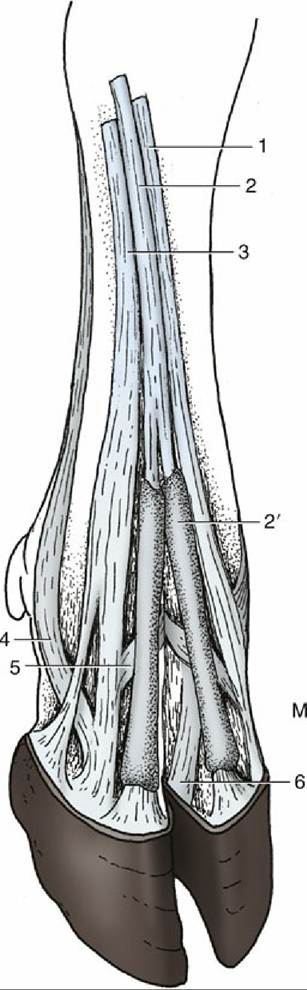
FIG. 30.9 Dorsal view of the bovine right forefoot. L, Lateral; M, medial; 1, medial tendon of common digital extensor to the medial digit; 2 and 2', common digital extensor and its sheaths, respectively; 3, lateral digital extensor; 4 and 5, abaxial and axial extensor branches, respectively, of the interosseous to the lateral digital extensor; 6, common axial collateral ligament.
1278