THE INTERNAL EAR
The mechanical stimuli produced by sound and by the positional changes of the head are transformed into nerve impulses in the internal ear. This is a delicate mechanism, no larger than about 12 mm across in the dog, and is completely enclosed in the very hard petrous temporal bone for protection and proper functioning (Figure 9-24, A).
It is exposed to sound vibrations on the lateral surface, and the impulses into which these are converted leave the bone in nerve fibers that pass through the internal acoustic meatus on the medial surface.The internal ear consists of a closed system of tiny membranous ducts and cavities known, because of its complexity, as the membranous labyrinth (Figure 9-28, A). This contains endolymph whose movement inside the system stimulates sensory cells in the membranous wall. Two enlargements in the center of the membranous labyrinth are known as the utriculus and sacculus. From the utriculus arise three semicircular ducts concerned with balance, and from the sacculus, the spiral cochlear duct, which is concerned with hearing.
The semicircular ducts stand roughly at right angles to each other and are designated anterior, posterior, and lateral; one end of each duct is ampullated close to the utriculus. The endolymph within them is set in motion by movements of the head, and this results in pressure on minute barriers (cristae ampullares) present in each ampulla (Figure 9-28/9,10). These pressures deflect sensory hairs projecting from the receptor cells of the cristae, stimulating the individual cells to send impulses to the central nervous system.
Two further receptor areas called maculae (Figure 9-28/6,7) are present in the walls of the utriculus and sacculus. They monitor the position of the head with respect to gravity. Although the maculae are bathed in endolymph, they react to a layer of crystals (statoconia) adhering to a gelatinous layer that surrounds the sensory hairs of the receptor cells.
When the gelatinous layer of the maculae faces toward the ground, the cells are maximally stimulated by the gravitational pull. The maculae thus record the position of the head, whereas the cristae record the movements of the head.The sacculus gives origin to the endolymphatic duct, which ends blindly in the epidural space (Figure 9-24/17). It is thought to function in the resorption of the endolymph secreted by the epithelial lining of the membranous labyrinth.
The membranous labyrinth is housed in a similar but slightly larger osseous labyrinth, a complex excavation in the temporal bone. The central chamber of the osseous labyrinth, the vestibule, houses the utriculus and the sacculus. The semicircular ducts lie within the osseous semicircular canals. The cochlear duct passes up the spiral canal of the cochlea, which is an excavation very similar to the inside of a snail’s shell. The center of the cochlea is an osseous pyramid known as
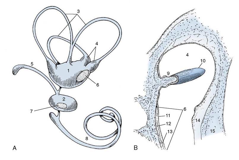
Figure 9-28 A, Membranous labyrinth. B, Section of ampulla. 1, Utriculus; 2, sacculus; 3, semicircular ducts; 4, ampullae containing ampullary crests; 5, endolymphatic duct; 6, 7, maculae; 8, cochlear duct; 9, ampullary crest; 10, cupula containing sensory hairs; 11, layer of neuroepithelial hair cells; 12, statoconia; 13, gelatinous layer of macula; 14, perilymphatic space; 15, wall of osseous labyrinth.
the modiolus (Figure 9-29/2). Running around the modiolus is the spiral canal, the actual lumen of the cochlea, which ends blindly at the apex of the modiolus. Projecting into the spiral canal from the modiolus is an osseous shelf, the spiral lamina (Figure 9-29/5), which terminates in the blind end of the spiral canal of the cochlea. The spiral lamina itself is hollow, forming the spiral canal of the modiolus.
Because the osseous labyrinth is slightly larger than the membranous labyrinth it encloses, there is a minute space between the two containing perilymph.
Only the two perilymphatic spaces (scala tympani and scala vestibuli) accompanying the cochlear duct into the cochlea need be considered further.The spiral canal of the cochlea is divided by a split longitudinal membrane into three channels (9-29/6,7,8), all running around the modiolus to the apex of the cochlea. The membrane arises centrally from the spiral lamina and, after splitting, attaches to the outside wall of the spiral canal. The uppermost channel is the scala vestibuli, the middle one is the cochlear duct, and the lowest is the scala tympani. The two scalae communicate at the apex of the cochlea around the blind end of the cochlear duct. At the base of the cochlea, the scala vestibuli communicates with the perilymphatic space in the vestibule, and the scala tympani ends at the secondary tympanic membrane (see Figure 9-24).
An enlarged transverse section of the spiral canal of the cochlea shows the composition of the split membrane, particularly the part that forms the walls of the triangular cochlear duct (Figure 9-29/7). The simplest of these walls separates the cochlear duct from the scala vestibuli; it consists of a single layer of cells and is known as the spiral membrane. The wall of the cochlear duct facing the scala tympani is complex by virtue of the large neuroreceptor and other cells found in it. Its connective tissue component is the basilar lamina, which plays an important role in the perception of sound. The cells form the spiral organ (Figure 9-29/13) in which originate the nerve impulses that are produced by the sounds received by the external ear.
The impulses travel toward the modiolus to ganglion cells housed in the spiral canal. The aggregate of these cells forms the spiral ganglion (Figure 9-29/14), which also winds around the modiolus. From the spiral ganglion the impulses travel along nerve fibers within canals to the base of the modiolus, where the fibers join to form the cochlear part of the vestibulocochlear nerve.
As the base of the stapes rocks in the vestibular window in unison with the vibrations of the tympanic
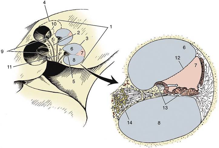
Figure 9-29 Cochlea and enlarged cochlear duct. 1, Cochlea; 2, modiolus; 3, 4, spiral canal of cochlea; 5, osseous spiral lamina; 6, scala vestibuli; 7, cochlear duct; 8, scala tympani; 9, 10, spiral canal of modiolus; 11, longitudinal canals; 12, spiral membrane; 13, spiral organ; 14, spiral ganglion.
membrane, it compresses the perilymph in the closed system of perilymphatic spaces. Because fluids are incompressible, the required “give” is found in similar vibrations of the secondary tympanic membrane closing the cochlear window. The way in which the mechanical stimuli in the vibrating columns of fluid within the cochlea act on the receptor cells in the spiral organ is complex and beyond the scope of this book. The width and structure of the basilar lamina suggests that, at least in humans, lower pitched sound is “read” by a relatively short stretch of the spiral organ near the apex of the cochlea. The remaining much longer stretch of the spiral organ responds to sounds of higher frequency, including those of speech.
The anatomy of the internal and middle ear is complicated by the passage of the facial nerve through this area (Figure 9-24/70). The facial nerve enters the internal acoustic meatus together with the vestibulocochlear nerve and, within an osseous facial canal, traverses the temporal bone to emerge at the stylomastoid foramen. The facial canal makes a sharp kneelike bend within the temporal bone, and at this point the nerve is enlarged by the geniculate ganglion. From this arises the major petrosal nerve, which regulates secretion of the lacrimal and nasal glands. The chorda tympani, regulating the sublingual and mandibular glands but also relaying taste from the rostral two thirds of the tongue, leaves the facial nerve a little more distally. The chorda tympani is so named because, for a short segment of its course, it lies on the upper part of the tympanic membrane (Figure 9-26/5). Both major petrosal and chorda tympani nerves leave the temporal bone through foramina on the rostroventral aspect of the bone. The facial nerve also supplies the stapedius muscle. (The tensor tympani is activated through the mandibular division of the trigeminal nerve [V3].)
The vestibulocochlear nerve (VIII) divides into vestibular and cochlear parts as it enters the internal acoustic meatus. The branches of the vestibular part pass to the neuroreceptor areas in the utriculus and sacculus, conveying impulses concerned with balance; the cochlear part passes into the base of the cochlea to mediate the impulses concerned with hearing.