THE INTESTINES
The intestines lie almost entirely to the right of the midline, packed mainly into the dorsal part of the abdomen and in part lying under cover of the ribs. Although said to measure as much as 50 m in adult cattle, their capacity is relatively slight, which is a feature correlated with the efficiency of gastric digestion.
Adhesion of the mesenteries of the small intestine and ascending colon during the fetal period results in these parts of the intestine sharing a common support in which they are flexed and coiled in a complex arrangement (see Figure 28-26) difficult to unravel in situ.The duodenum takes origin below the ribs. Its first part rises almost vertically toward the visceral surface of the liver; it then runs toward the pelvis as the descending duodenum but turns when almost level with the coxal tuber. The ascending part then returns toward the liver, passing to the left of the cranial mesenteric artery, to enter the fringe of the mesentery. It is continued by the jejunum. The first part of the duodenum is joined to the liver by the lesser omentum. The other border of the first and descending parts gives attachment, directly or at slight remove, to both walls of the greater omentum (Figures 28-4, C, and 28-24). Only the descending duodenum is immediately visible on opening the right flank.
The jejunum forms many short coils within the free margin of the mesentery. Their general course takes them ventrally, then caudally, and finally dorsally toward the large bowel. The position of these coils depends on the fullness of the rumen and the size of the uterus; usually most lie within the supraomental recess, but some may spill from this to insinuate themselves behind the rumen and so appear against the left flank. The extent of the short ileum is defined by the ileocecal fold (Figure 28-26/4,6).
The cecum continues into the colon without obvious change in diameter; the junction is marked only by the entrance of the ileum.
Its rounded blind tip projects caudally from the supraomental recess and floats high when gas-filled. When greatly distended with gas for protracted periods, it must be deflated surgically. Rotation of the cecum together with the proximal loop of the colon (Figure 28-26/7) is common, compromises its function and blood supply, and requires surgical correction.The colon is divided into the usual ascending, transverse, and descending parts (see Figure 3-45/Ru). The
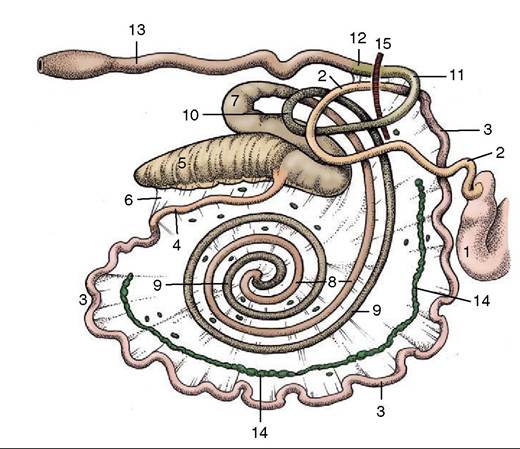
Figure 28-26 Right lateral view of the bovine intestinal tract, schematic. 1, Pyloric part of abomasum; 2, duodenum; 3, jejunum; 4, ileum; 5, cecum; 6, ileocecal fold; 7-10, ascending colon; 7, proximal loop of ascending colon; 8, centripetal turns of spiral colon; 9, centrifugal turns of spiral colon; 10, distal loop of ascending colon; 11, transverse colon; 12, descending colon; 13, rectum; 14, jejunal lymph nodes; 15, cranial mesenteric artery.
first of these is wound in a very elaborate manner. On leaving the cecum it forms a flattened sigmoid flexure (see Figure 3-45/11) before narrowing and turning ventrally to trace a double spiral attached to the left side of the mesentery. Two centripetal turns are succeeded by two centrifugal turns that restore the colon toward the periphery of the mesentery, where it continues into a distal loop that carries it first toward and then away from the pelvis (see Figure 3-45/11'). Beyond this it joins the short transverse colon that crosses the midline in front of the mesenteric artery and leads directly into the descending colon. This part runs toward the pelvic entrance within a mesentery that is thickened by fat and fused with neighboring parts of the gut. The mesentery of the descending colon is at first short but lengthens in front of the sacrum, where the colon forms a sigmoid flexure before continuing as the rectum. This looseness gives the hand of the veterinarian considerable range in rectal exploration (p.
720). The rectum is described with the pelvic viscera.The ascending colon of small ruminants performs three or four turns in each direction. A more significant difference lies in the “pearl necklace” appearance of the centrifugal turns, in which the contents are already segmented into the pellets so characteristic of the feces. The string of these pellets in the ascending colon is replaced by their massing in a thicker column in the wider descending colon and rectum.
Few features of the interior of the intestines call for comment. In cattle the accessory pancreatic duct opens far down the descending duodenal limb, the bile duct opens more proximally, where the duodenum lies against the liver. In the small ruminants the greater pancreatic duct is usually present. The ileum projects into the cecum, and a low rampart is thus present around the ileal orifice. Lymphoid tissue is generously spread through the mucosa, especially in the small intestine, where both solitary and aggregated nodules occur. The aggregated nodules may reach lengths of 25 cm and are distinguished by their irregular cribriform surfaces. Usually one of these patches extends through the ileal orifice into the large gut.
The bulk of the intestines is supplied by the cranial mesenteric artery; however, the first part of the duodenum is supplied from the celiac artery and the descending colon is supplied from the caudal mesenteric artery. The intestinal veins combine to form the cranial mesenteric radicle of the portal vein. Many jejunal lymph nodes are found within the mesentery, where they form a more or less continuous chain of giant nodes placed between the peripheral festoons of small intestine and the more central coils of the spiral colon (Figure 2826/14). The largest may be as much as a meter in length. In the small ruminants this chain of nodes lies central to the last centrifugal turn of the spiral colon. Other small nodes are scattered beside the cecum, colon, and rectum. The efferent stream from the mesenteric nodes joins the cisterna chyli. The nerves that reach the gut along the cranial mesenteric artery consist of both sympathetic and vagal fibers. The parasympathetic nerves to the last part of the colon are derived from the sacral outflow.