POSTNATAL DEVELOPMENT
At birth the ruminant stomach is prepared for the digestion of milk. The abomasum predominates and is remarkable not only for its size, which surpasses the combined capacity of the other chambers, but also for the degree of structural maturity that it has attained.
Its full extent is apparent directly after the consumption of a generous feed, when it extends from the liver and diaphragm to the pelvic entrance, from one flank to the other, and from the floor well into the upper half of the abdomen (Figures 28-20, A, and 28-25/4). Its capacity may already exceed 60% of the adult measure. So large an organ inevitably impinges on almost all other abdominal contents, but only the extensive contact with the liver, which in the neonate reaches far across the median plane, need be mentioned. The abomasal mucosa is at first not quite mature, and a few days elapse before the fundic glands become fully active; presumably this is a provision to allow the absorption of unaltered antibodies from the colostrum.In contrast to the abomasum, the rumen and reticulum of the newborn calf are very small. They are confined to the left dorsal and cranial corner of the abdomen and are generally found crumpled and collapsed (Figure
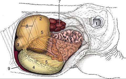
Figure 28-25 Topography of the abdominal organs in a newborn calf, left lateral view. The left abdominal wall and the left hindlimb have been removed. 1, Left acetabulum; 2, rumen; 3, reticulum; 4, abomasum; 5, greater omentum; 6, small intestine; 7, left kidney; 8, position of spleen; 9, liver.
28-25/2,5); they are bypassed by milk feeds and normally contain only a small amount of fluid—secretions of the respiratory tract (swallowed in utero) in the youngest animals and saliva in those a little older. The omasum is also retarded in development and forms a relatively inconspicuous bridge between the reticulum and the abomasal fundus.
The walls of the forechambers are thin and deficient in muscle, and while their mucosae possess the characteristic adult features, these are present in subdued form.No striking changes in proportions and structure are to be observed before the young calf shows serious interest in solid food, generally from the time it is 2 or 3 weeks old. Thereafter the abomasum continues to increase at a slow but steady rate while the rumen and reticulum enter a period of spectacular growth. They have generally overtaken the abomasum by 8 weeks, and at 12 weeks they are more than twice as large. This unequal growth continues—but more slowly—until the time when the definitive topography and proportions are established. It is difficult to specify this age, for many variable factors are involved; some authors assert that the conformation is virtually adult after 3 months, but others believe that it does not become so until near the end of the first year.
Normal development depends on the availability of a normal diet of solid forage, but there remain some uncertainties concerning the precise stimuli that are involved. At one time it was thought that roughage not only stretched the stomach wall and stimulated its muscular growth but also promoted the differentiation of the mucosa. Later it was shown that many gross and microscopic features of the mucosa develop only with exposure to certain end products of microbial fermentation, notably butyric acid. Exposure to these stimuli must be continued for some time if development is to follow its normal course, and the return of a young, partly weaned calf to a wholly milk diet may result in the arrest and sometimes even reversal of the maturation processes.
The abomasum is initially the most vigorous chamber, but its activity diminishes as the raminoreticulum, first inert and then only spasmodically active, establishes a regular cycle of contraction by the second month. The feeding habits, the structural changes, and the motor and chemical activities of the stomach, when taken in conjunction, define three phases of development.
A neonatal period, in which milk forms the sole diet, may last for 2 or at most 3 weeks and may be followed by a transitional period when the stomach is adapting to solid food. From the eighth week onward the anatomy and the processes of digestion may be essentially those of the adult. The chronology will clearly be different in dairy and suckler calves.Changes in abdominal topography are not confined to the stomach. In the newborn the liver is relatively large and lies across the midline, extensively related to the abomasum. As the rumen and reticulum increase in size the liver is pressed toward the right and dorsally, and it rotates so that its left lobe comes to lie cranio- ventral to the right one and out of the reach of the abomasum. The intestines are simultaneously pushed away from the left flank and become confined to the right side; the expansion of the dorsal ruminal sac also displaces the left kidney, thrusting it across the midline until it comes to rest below and caudal to its fellow (Figures 28-11/9 and 29-9/29).