THE KIDNEYS
The account of the kidneys presented here concentrates on their positions and relations. Other aspects of their anatomy are considered in Chapter 15.
The kidneys in the dog are bean-shaped and retro- peritoneally positioned against the sublumbar muscles.
The right kidney is usually said to lie below the first three lumbar vertebrae, and the left one is said to lie below the second to fourth (Figure 14-22); however, this may specify their positions too definitely and they may be found a full vertebral length more caudally. The right kidney is more restricted by being deeply recessed within the liver and is related medially to the right adrenal gland and caudal vena cava, laterally to the last rib and abdominal wall, and ventrally to the liver and pancreas (Figure 14-23). The left kidney is related cranially to the spleen (or stomach when enlarged), medially to the left adrenal gland and aorta, laterally to the abdominal wall, and ventrally to the descending colon.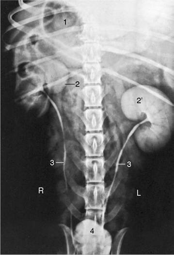
Figure 14-22 Urogram of a dog. 1, Gas in stomach; 2, 2', right and left kidneys; 3, ureters; 4, bladder.
The cat’s kidneys are relatively large and are given a distinctive appearance by capsular veins converging over the surface toward the hilus (Figure 14-24). They are more mobile than the kidneys of the dog (see Figures 14-13 and 14-14), especially the left one, which can be displaced cranially or caudally from its usual position below the second to fifth lumbar vertebrae; it has been taken for a pathological swelling. In cats, both kidneys are readily palpable.