THE KIDNEYS
The positions and relations of the kidneys were described in the previous chapter.
The right kidney usually lies below the first three lumbar vertebrae, and the left one lies below the second to fourth, although both may be found a full vertebral length more caudally.
In the bitch the caudal poles of both kidneys reach close to, or make contact with, the fat-filled mesovaria. Although described as unipyrami- dal (p. 177), the canine kidney retains clear evidence of the former existence of a number of separate pyramids. The renal arteries, direct branches from the aorta, usually divide before entering the kidneys; they may be assisted by small arteries of aberrant origin. The renal veins pass directly to the caudal vena cava (Figure 14-21). There are no features of major specific interest in the sympathetic and parasympathetic nerve supply.The kidneys of the cat are relatively larger, shorter, and thicker than those of the dog and obtain a distinctive appearance from the capsular veins that converge toward the hilus, where they enter the renal vein (Figure 15-5). The cut surface of the kidney is red to yellowish red because of a large amount of intracellular fat stored in the proximal convoluted tubules; the fat content is greatest in castrated males and pregnant females. There are fewer vestiges of the multipyramidal stage of development. The kidneys are more mobile than in dogs,
2
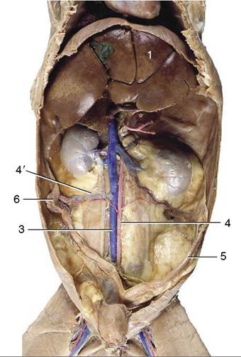
Figure 15-5 Ventral view of feline abdominal roof. 1, Liver; 2, kidneys (with stellate v.v.); 3, caudal vena cava (injected);
4, aorta; 4', ovarian a. (injected); 5, uterine horn; 6, ovary.
especially the left one, which can be displaced rather far cranially or caudally from its usual position (Figure 14-13); it has been mistaken for a pathological swelling.
In cats, both kidneys are readily palpable.In the dog (if not the cat) it is generally thought more prudent to expose a kidney by laparotomy when a biopsy specimen is required rather than to attempt a blind puncture.
The muscle of the renal pelvis is strongest at the transition to the ureter, presumably to impel urine into the narrower tube. The abdominal part of the ureter runs retroperitoneally close to the aorta or vena cava (Figures 14-21, 14-22/5, and 15-5), passing over the dorsal (lateral) surface of the gonadal vessels before crossing the ventral face of the deep circumflex iliac vessels and the terminal branches of the aorta (and corresponding veins). It is carried into the pelvis in the base of the broad ligament or genital fold, which brings it to the dorsal surface of the bladder; in the male it crosses above the deferent duct toward the end of its course. It penetrates the bladder wall very obliquely. The inclusion of the ureter within the genital fold places it at some risk in the common spay operation.
Survey radiographs of the abdomen will adequately reveal the external anatomy of the kidneys when, as is usually the case, they are enclosed in fat. (Deficiency of fat occurs in very young pups and in emaciated older
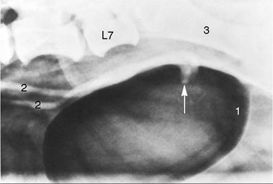
Figure 15-6 The canine bladder made visible by the introduction of air. The arrow indicates the terminations of the ureters in the dorsal wall of the bladder, superimposed here on the air-filled lumen. 1, Caudal end of bladder; 2, ureters; 3, shaft of ilium.
subjects.) Visualization of internal features requires the intravenous injection of an appropriate contrast agent that is then excreted in the urine; suitably staged radiographs will show general opacification of the cortex and medulla (Figure 14-22), renal pelvic morphology (Figure 5-29), and, later, the status of the ureters and bladder. Because the passage of urine is assisted by peristaltic contraction, a single radiograph does not usually depict a healthy ureter along its entire length.