The Kidneys
The shape of the pig's kidneys is very distinctive. They are flattened (see Fig. 5.21C) against the abdominal roof (within a fatty capsule), extending from the level of the last rib to that of the fourth lumbar vertebra (Fig.
34.15/5). This symmetry of position is most unusual and deprives the right kidney of the expected contact with the liver. The left kidney is related ventrally to the colic spiral, the cecum, and the pancreas; the right one is related to the descending duodenum and also possibly to the pancreas.The internal structure resembles that of the human kidney (Fig. 34.16). A central cavity with two recesses (major calices) directed toward the poles comprises the pelvis, which extends about a dozen minor calices, each embracing a renal papilla through which the papillary ducts discharge urine. The papillae correspond to renal pyramids, and because the number of these is reduced by fusions in the course of development, there is some inequality in the size of the units presented by the mature organ.
'" 7
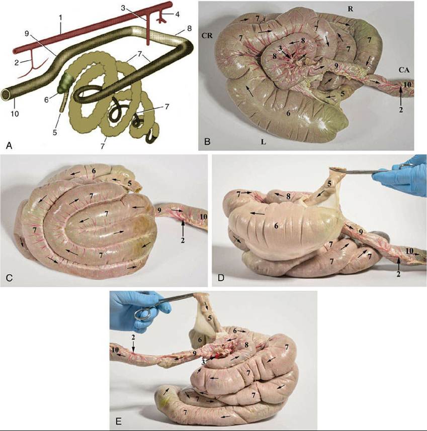
FIG. 34.12 The large intestine, schematic view from the right side (A), dorsal view (B), ventral view (C), left side (D), and right side (E). 1, Aorta; 2, caudal mesenteric artery; 3, cranial mesenteric artery; 4, celiac artery; 5, ileum; 6, cecum; 7, ascending colon; 8, transverse colon; 9, descending colon; 10, rectum.
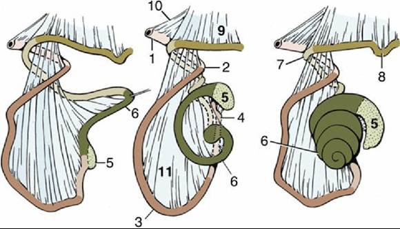
FIG. 34.13 The development of the ascending colon, left lateral view. 1, Descending duodenum; 2, caudal flexure of duodenum; 3, jejunum; 4, ileum; 5, cecum; 6, ascending colon; 7, transverse colon; 8,
descending colon; 9, descending mesocolon; 10, mesoduodenum; 11, mesentery.
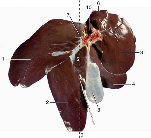
FIG. 34.14 Visceral surface of the liver. 1, Left lateral lobe; 2, left medial lobe; 3, right lateral lobe; 4, right medial lobe; 5, quadrate lobe; 6, caudate process; 7, porta; 8, gallbladder; 9, approximate position of median plane; 10, caudal vena cava.
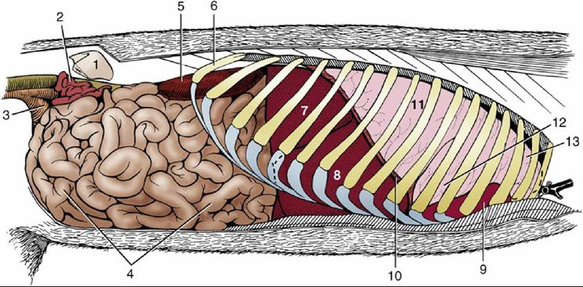
FIG. 34.15 Abdominal and thoracic viscera, right lateral view. 1, Wing of ilium; 2, uterine horns; 3, bladder; 4, jejunum; 5, right kidney; 6, last rib; 7 and 8, right lateral and medial lobes of liver; 9, heart in pericardium; 10, diaphragm, cut; 11-13, caudal, middle, and cranial lobes of right lung.