» The Kidneys and Adrenal Glands
The kidneys of adult cattle retain much of their fetal Iobation and are divided by surface fissures into about a dozen lobes (see Figs. 5.21 and 5.23). The right kidney has a flattened ellipsoidal form and lies in a conventional position with a dorsal retroperitoneal attachment to the sublumbar musculature.
It is received cranially into the renal impression of the liver. The left kidney is less regular, being flattened at its cranial pole and thickened caudally. Its position below and behind its fellow is unusual and is the consequence of the postnatal growth of the rumen (see Fig. 29.9/10). Although surrounded by considerable accumulations of fat (capsula adiposa), both kidneys vary in position with the phase of respiration and according to the pressure exerted by other viscera. In the cadaver the right kidney is commonly found below the last rib and first two or three lumbar transverse processes, while the left one lies at a more ventral level under the second to fourth lumbar vertebrae. The left kidney is accessible on rectal exploration, but the right one usually is not. The left kidney may return to the left side when the pressure on it is relieved by fasting in life or after evisceration in the course of an autopsy.The numerous relations of the right kidney need not be described at length. They include the liver, pancreas, duodenum, colon, and, in most animals, the adrenal gland. The hilus is widely open and lies ventromedially; the ureter runs from it, crossing the medial margin to follow a winding retroperitoneal course below the abdominal roof that carries it into the pelvis.
The left kidney is swung through about 90 degrees around the axis of the aorta in moving from its fetal (see Fig. 28.25) to its adult location against the right face of the dorsal sac. It hangs in a relatively long fold, rests on the intestinal mass, and is flattened by contact with the rumen.
The left ureter crosses the dorsal aspect of the kidney to regain the left half of the abdomen. Its later course is similar to that of the right duct.In structure the bovine kidneys are of the multipyramidal type (Fig. 28.27). The separate medullary pyramids are capped by a continuous cortex, which appears fragmented by fissures extending inward from its surface (Fig. 28.28). The cortex (Fig. 28.27/4) is clothed in a tough capsule that is easily stripped from the healthy organ, except toward the hilus, where it blends with the wall of the ureter. The cortical and medullary regions are distinguishable in gross sections by the much lighter color of the former and by the cut vessels that mark their mutual boundary. The glomerular vascular tufts scattered through the cortex may be visible to the naked eye. The apex (papilla; Fig. 28.27/3) of each medullary pyramid fits into a calyx or cup formed by one of the terminal branches of the ureter; these branches eventually unite to form two major channels that converge from the cranial and caudal poles to yield a single ureter (see Fig. 5.23). There is thus no large central expansion corresponding to a renal pelvis.
The renal arteries are derived from the aorta; the renal veins join the caudal vena cava. Lymphatic vessels lead to the renal nodes, enlarged members of the lumbar aortic series, and these in turn drain into the lumbar lymph trunk.
The kidneys of the sheep and goat are quite unlike those of cattle but conform closely in external appearance and internal structure to those of the dog (see Fig. 5.23). They are more regular in shape than the dog's, being protected from distorting pressures by enclosure in thick masses of fat. The fat cushion makes the left kidney less subject to displacement by the rumen.

FIG. 28.27 Bovine kidney dissected to show its interior (semi-schematic). 1, Principal branches of ureter;
2, calyx; 3, renal papillae; 4, renal cortex; 5, interlobular artery.
The adrenal glands are located close to the kidneys. The right gland is heart shaped and usually lies against the medial margin of the cranial extremity of the corresponding kidney (Fig. 28.10/12). The left one is less regular in form and less constant in position; generally it is found within the perirenal fat some centimeters cranial to the left kidney. The division into cortex and medulla is very evident in gross sections.
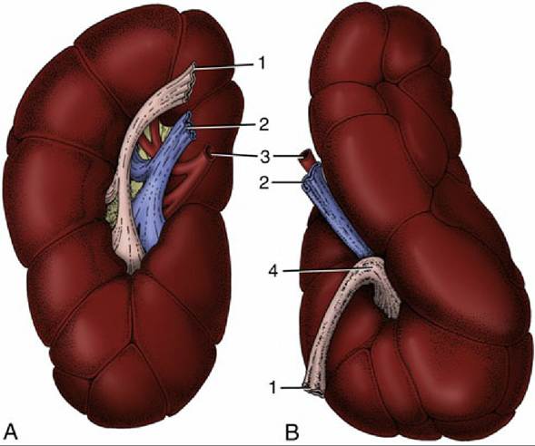
FIG. 28.28 Ventral views of the (A) right and (B) left bovine kidneys. 1, Ureter; 2, renal vein; 3, renal artery; 4, renal sinus.
The Lymph Nodes of the Abdominal Roof
A number of important lymph nodes are scattered about the bifurcation of the aorta and between its terminal branches. Most belong to the medial iliac group, which collects lymph from the hindlimbs, pelvic walls, and pelvic viscera (see Fig. 29.4). The large deep inguinal (iliofemoral) node, in the angle between the external and deep circumflex iliac arteries, receives the flow from the udder; when enlarged it can be palpated per rectum near the cranial border of the ilium. The efferent stream forms the lumbar trunk, which runs forward over the aorta to enter the cisterna chyli. A few much smaller (lumbar aortic) nodes that are spread along the psoas musculature are concerned with the lymphatic drainage of the vertebrae and neighboring muscles. The renal nodes belong to this series.
Comprehension Check
With the aid of diagrams, explain the anatomic relationships that arise in the bovine abdomen when the abomasum is normally situated, when it is displaced to the left, and when it is displaced to the right.
Review the innervation of the abdominal wall and the paravertebral nerve blocks to anesthetize the abdominal wall for laparotomy.
>-------------------- <
* In wild ruminants, striking changes in the total mass of the salivary glands are correlated with the ruminal response to the fibrous content of the forage.
* There is evidence that the infective (prion protein) agents responsible for the transmissible spongiform encephalopathies (e.g., bovine spongiform encephalopathy) reach the central nervous system by transport from the gut along the splanchnic and vagal nerves.