» The Pelvic Cavity
The pelvic cavity of the cow becomes progressively narrower between the entrance and the exit. There is a pronounced dip of the middle part of the floor resulting in a local increase in height before the caudal part slopes steeply upward to the shallow exit (Fig.
29.1).The entrance faces ventrocranially at an angle that carries the pecten of the pubis below the second intersacral joint (Fig. 29.1/15). Behind the iliac shaft, the width is reduced by inflection of the high ischial spine, and it becomes further reduced by the encroachment of the massive ischial tuber on the exit (Fig. 29.2). The conspicuously cramped exit is roughly triangular; the third caudal vertebra and the tubercular ischial tubers are its corners. The lateral border is completed by the sacrotuberous ligament (the edge of the sacrosciatic ligament), while the caudal margin of the floor is cut away at the ischial arch. The strong development of the ischial crest and tuber combine to reduce the contribution to the lateral wall that is made by the sacrosciatic ligament (Fig. 29.2/4).
There are certain variations associated with age and gender. The entrance is almost uniformly wide in mature cows but considerably narrowed in its ventral part in heifers. In these younger animals the cranial part of the floor raises a ridge over the symphysis; in older cows, especially those that have carried several calves, the same region is level or sunken. The male girdle, despite being significantly more robust, encloses a cavity that is clearly less capacious. It is even more confined at the entrance, and beyond this the cranial part of the floor tends to be domed.
In sheep and goats the long, slender iliac shafts approach the vertebral column at an acute angle that, in combination with the shortness of the sacrum, places the pecten below the second joint of the tail (see Fig.
26.2).The sacroiliac joints (Fig. 29.3) are complemented and rendered virtually immobile by strong ligaments between the two bones. About the time of parturition, hormones induce some slackening of the collagenous structures of the pelvis to allow modest but potentially significant mobility (p. 200). Ankylosis of these joints, accompanied by lumbar spondylosis, is common in aging bulls and, when severe, may disable the animal for service.
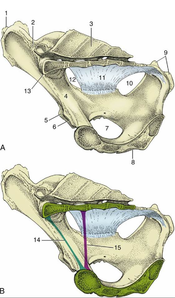
FIG. 29.1 (A) and (B), Median section of the bony pelvis of a cow. Certain obstetrical terms are illustrated in (B). 1, Coxal tuber; 2, sacroiliac joint; 3, sacrum; 4, shaft of ilium; 5, cranial border of acetabulum; 6, pecten pubis; 7, obturator foramen; 8, symphysis; 9, ischial tuber; 10, lesser sciatic foramen; 11, sacrosciatic ligament; 12, greater sciatic foramen; 13, promontory; 14, conjugate (the line connecting the promontory with the pecten); 15, vertical diameter (the vertical line between the pectin and the pelvic roof).
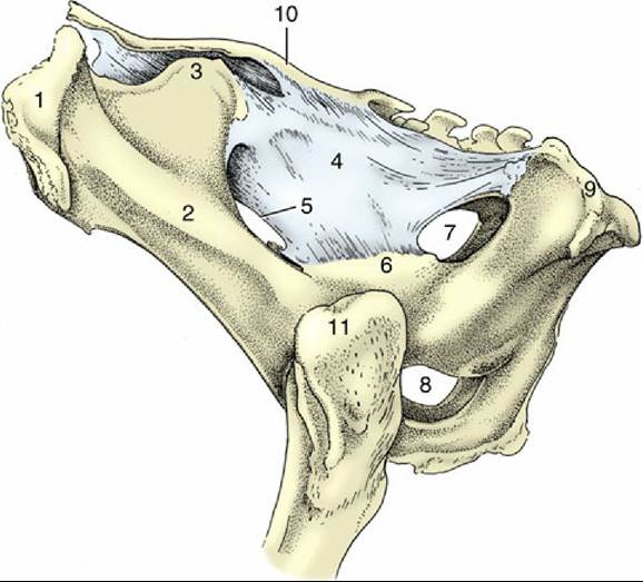
FIG. 29.2 Lateral view of the bony pelvis of a cow. 1, Coxal tuber; 2, shaft of ilium; 3, sacral tuber; 4, sacrosciatic ligament; 5, greater sciatic foramen; 6, ischial spine; 7, lesser sciatic foramen; 8, right and left obturator foramina; 9, ischial tuber; 10, sacrum; 11, greater trochanter.
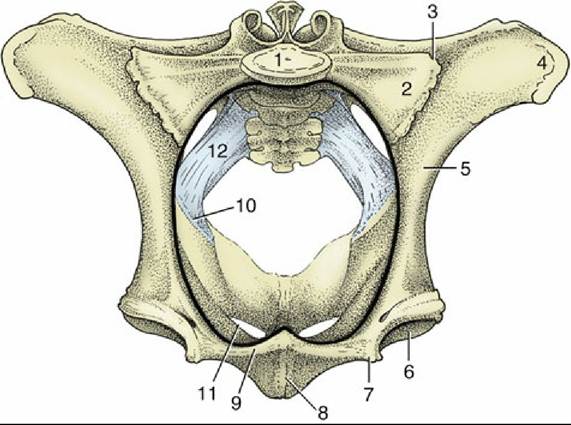
FIG. 29.3 Cranial view of the bony pelvis of a cow. The terminal line (black) is indicated. 1, Body of first sacral vertebra; 2, wing of sacrum; 3, sacroiliac joint; 4, coxal tuber; 5, shaft of ilium; 6, acetabulum; 7, iliopubic eminence; 8, symphysis; 9, pecten pubis; 10, ischial spine; 11, obturator foramen; 12, sacrosciatic ligament.
The perineal region is extensive because those parts of the hamstring musculature that in the horse provide it with very prominent lateral boundaries are lacking in cattle.
By convention, the region is considered to extend ventrally to include the nearest part of the udder (or scrotum). The increase in breadth exposes the sacrotuberous ligaments, the ischial tubers, and the ischiorectal fossae as visible and palpable surface landmarks. The anus and vulva, the most obvious features of the dorsal and ventral perineal regions, respectively, are considered later (see Fig. 29.10).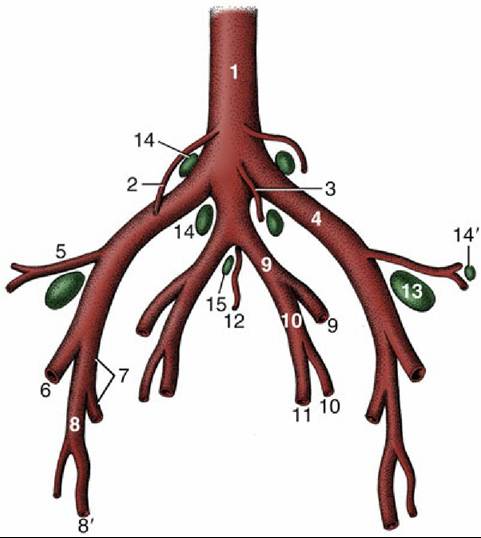
FIG. 29.4 Branching pattern of the caudal part of the bovine abdominal aorta. 1, Aorta; 2, ovarian artery; 3, caudal mesenteric artery; 4, external iliac artery; 5, deep circumflex iliac artery; 6, femoral artery; 7, deep femoral artery; 8, pudendoepigastric trunk; 8', external pudendal artery; 9, internal iliac artery; 10, umbilical artery; 11, uterine artery; 12, median sacral artery; l3, deep inguinal (iliofemoral) lymph node; 14 and 14', medial and lateral iliac lymph nodes, respectively; 15, sacral lymph nodes.
The blood supply to pelvic structures is delivered by the small median sacral artery and the much larger, paired internal iliacs (Fig. 29.4). The first or, more accurately, its continuation as the median caudal artery has already been encountered (p. 656). The internal iliac artery serves both parietal and visceral structures, contrary to the usual arrangement. It enters the pelvic cavity close to the sacroiliac joint and continues down the ilium to reach the vicinity of the lesser sciatic foramen (Fig. 29.1/10) before dividing into internal pudendal and caudal gluteal arteries. The latter, like other parietal branches, is of no present concern. The internal iliac's first visceral branch, detached close to the origin of the parent trunk, is the umbilical artery. This term, though appropriate to its role in the fetus, is misleading because the vessel is now almost exclusively concerned with supplying blood to the uterus through a large uterine artery; the continuation of the umbilical, reduced to a fibrous cord with a vestigial lumen, is better known as the round ligament of the bladder.
The male homologue of the uterine artery is the deferential. (The distribution of the arteries to the viscera is considered with the organs they supply.) The second visceral branch, the vaginal artery, is detached close to the termination of the internal iliac trunk and supplies the bulk of the pelvic viscera. The male homologue is the prostatic artery. The internal pudendal artery supplies both parietal structures, including the muscles of the pelvic diaphragm, and viscera, including the female tract from the caudal vagina to the vestibule. The depleted trunk leaves the pelvis, through an opening in the fascia directly above the symphysis, to supply branches to the clitoris and labia and other branches to the perineum, some of which reach the caudal part of the udder (or scrotum and prepuce).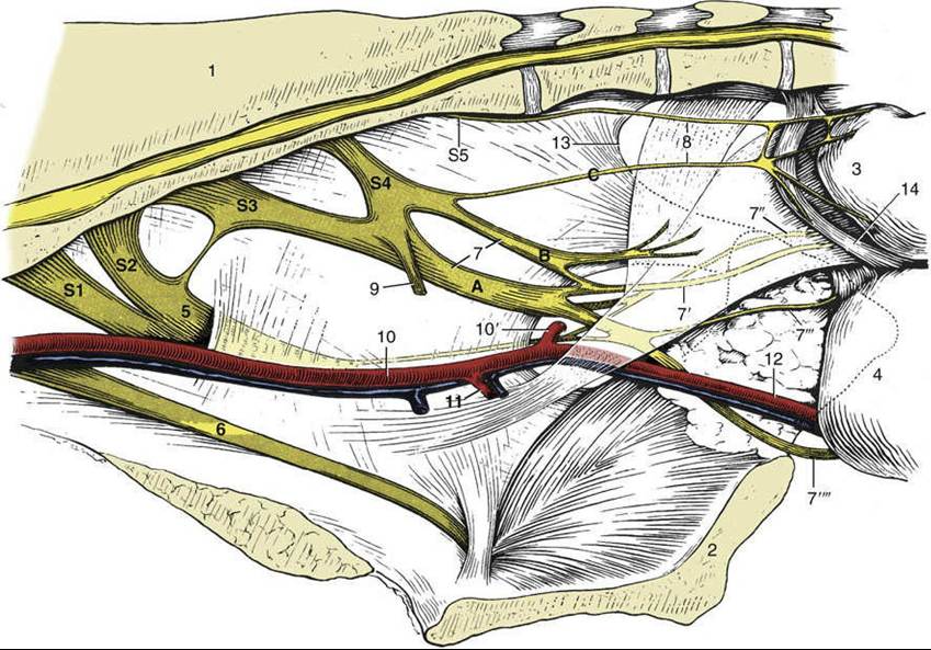
FIG. 29.5 Nerves and vessels on the medial surface of the bovine pelvic wall. Local anesthesia of the pudendal nerve (n.) can be obtained by injections at points A and B; anesthesia of the caudal rectal nerves is possible by an injection at point C. 1, Sacrum; 2, pelvic symphysis; 3, rectum (reflected); 4, vagina (reflected); 5, sciatic n.; 6, obturator n.; 7, pudendal n.; 7', distal cutaneous branch of pudendal n.;
7", proximal cutaneous branch of pudendal n.; 7'", deep perineal n.; 7"", continuation of pudendal n. to clitoris; 8, caudal rectal nerves; 9, pelvic n.; 10, internal iliac artery (a.); 10', caudal gluteal a.; 11, vaginal a.; 12, internal pudendal a.; 13, caudal border of sacrosciatic ligament; 14, retractor clitoridis; S1 to S5, sacral nerves 1 through 5.
The nerves within the pelvis fall into two groups (Fig. 29.5). The first comprises the obturator and sciatic nerves that, despite their vulnerability to injury at parturition, will be described with the hindlimb. The second group comprises the pudendal, caudal rectal, and pelvic nerves, of which all are purely sacral in origin and concerned with the supply of the pelvic viscera and the perineum.
The significant divisions of the pudendal nerve are the deep perineal and distal cutaneous branches and the continuation of the main trunk. The deep perineal supplies both visceral and somatic structures of the caudal pelvic region. The distal cutaneous branch supplies structures of the ventral perineum (before it becomes superficial by emerging from the ischiorectal fossa), crosses the medial process of the ischial tuber (where it may be palpated), and supplies the vulva and perineal skin; some branches extend as far as the nearest part of the udder. The depleted trunk passes ventral and leaves the pelvis in company with the internal pudendal artery; it supplies the dorsal nerve of the clitoris/penis and supplies other branches to the skin of the udder/scrotum and prepuce.Pudendal nerve block is used in surgery of the prepuce in bulls and also in the management of chronic prolapse in cows. The block is instituted by locating the lesser sciatic foramen and the nerve that lies on the sacrosciatic ligament a bit craniodorsal to the foramen. The needle, inserted via the ischiorectal fossa, is guided close to the nerve followed by depositing of the anesthetic.
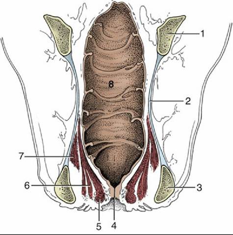
FIG. 29.6 Dorsal section of the bovine rectum and adjacent structures. Note especially the topography of the pelvic diaphragm (6 and 7). 1, Shaft of ilium; 2, sacrosciatic ligament; 3, ischial tuber; 4, anus; 5, external anal sphincter; 6, levator ani; 7, coccygeus; 8, rectum.