The Large Intestine
The large intestine is capacious and, like that of the horse, is much sacculated, being drawn into a series of pouches by two (on the colon) or three (on the cecum) taeniae that run along its length.
The peculiar disposition presented by the cecum and ascending colon in this animal, unique among domestic species, results from the greater than 360-degree rotation performed by the loop of bowel that is herniated into the umbilical cord early in development (see Figs. 3.64 and 3.65). This carries the caudal limb of the loop, including the cecocolic junction, to the left of the mesenteric axis, where it remains throughout later development and into adult life. The ascending colon thus commences on the left side and only gains its usual continuation into the transverse colon on the right side of the abdomen in consequence of the reversal of course described next.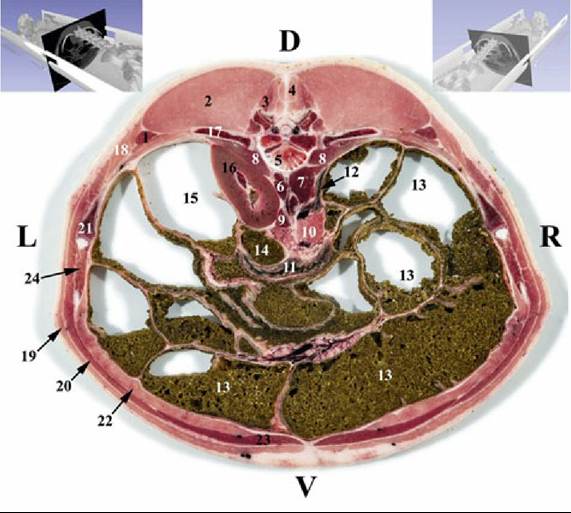
FIG. 34.11 Transverse section of the abdomen of the pig at the level of the 16th thoracic vertebra. 1, Iliocostalis muscle; 2, longissimus dorsi muscle; 3, multifidi muscles; 4, spinalis muscle; 5, 16th thoracic vertebra; 6, diaphragm, left crus; 7, diaphragm, right crus; 8, psoas minor muscle; 9, left adrenal gland; 10, pancreas, right lobe; 11, dudodenum, caudal duodenal flexure; 12, ascending duodenum; 13, ascending colon; 14, descending colon; 15, cecum; 16, left kidney; 17, 16th rib; 18, serratus dorsalis muscle, caudal part; 19, cutaneus trunci muscle; 20, obliquus externus abdominis muscle; 21, 15th rib; 22, transversus abdominis muscle; 23, rectus abdominis muscle; 24, intercartilaginei muscles.
The cecum and colon must be considered together because they combine in a conical, ventrally tapering mass suspended from the roof of the abdomen (Fig. 34.12). The cecum, which has a capacity of about 2 L, has its origin below the left kidney and extends ventrally or caudoventrally against the left flank to its rounded, blind apex.
The ascending colon is arranged around its mesentery in a cone that points ventrally to reach the abdominal floor (with some deviation possible in any direction) (Fig. 34.12). The outer part of the cone is provided by the wide, sacculated portion continuing from the cecum; when viewed from above, it spirals ventrally, clockwise and centripetally, before reversing course at the apex of the cone to ascend in narrower, smoother, and tighter centrifugal coils concealed within the center of the cone. These carry it dorsally to emerge from the base of the cone, pass to the right of the root of the mesentery, and continue as the transverse colon. The cecocolic mass mainly occupies the middle third of the left side of the abdomen, leaving the caudal and right regions available to the jejunum. However, variation is common and, especially where the jejunum is concerned, can be considerable. There is little notable about the remainder of the large intestine beyond the existence of a rectal ampulla.