The Small Intestine
The duodenum is also arranged rather like that of the dog, descending toward the pelvis before turning to run forward to the left of the root of the mesentery before dipping ventrally to be continued by the jejunum (Fig.
34.6 and 34.7). It is entered by the bile duct about 3 cm beyond the pylorus and by the single (accessory) pancreatic duct about 10 cm farther on. Both openings are raised on papillae.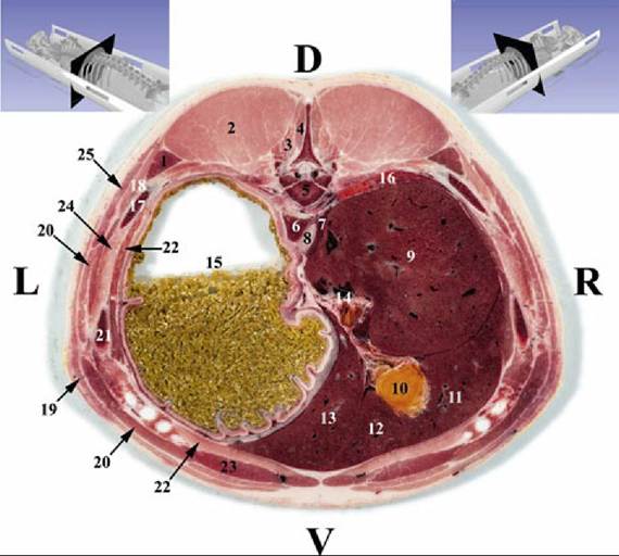
FIG. 34.10 Transverse section of the abdomen of the pig at the level of the 11th thoracic vertebra. 1, Iliocostalis thoracis muscle; 2, longissimus dorsi muscle; 3, multifidi muscles; 4, spinalis muscle; 5, 11th thoracic vertebra; 6, diaphragm, left crus; 7, diaphragm, right crus; 8, esophagus; 9, liver (right lateral lobe); 10, gall bladder; 11, liver (right medial lobe); 12, liver (quadrate lobe); 13, liver (left medial lobe); 14, porta hepatis; 15, stomach; 16, right lung, caudal lobe; 17, 11th rib; 18, serratus dorsalis muscle, caudal part; 19, cutaneus trunci muscle; 20, obliquus externus abdominis muscle; 21, 10th rib; 22, transversus abdominis muscle; 23, rectus abdominis muscle; 24, intercostalis muscles; 25, latissimus dorsi muscle.
The jejunum is arranged in many small loops (see Fig. 34.13) suspended by a mesentery that gives them much freedom of position. The greater part lies in the right half of the abdomen, ventrally and toward the pelvis, but some part may be in contact with the left flank behind the colic spiral. Like many other abdominal organs, the jejunum must accommodate its position to the condition of the stomach and, in sows, to that of the uterus.