The larynx is located caudal to the intermandibular space and ventral to the first two or three cervical vertebrae.
Its cranial parts can be examined through the mouth in the sedated dog when the soft palate is raised with a spatula (Fig. 11.31A). Palpation through the skin reveals, in caudorostral succession, the cricoid cartilage (especially its arch), the rounded ventral surface of the thyroid cartilage, and the prominent thyrohyoids that connect the rostral horns of the thyroid cartilage with the basihyoid.
The remaining bones of the hyoid apparatus, other than the stylohyoid, are also palpable (Figs. 2.34, 11.9, and 11.32).The epiglottis resembles a pointed spade that is connected to the body of the hyoid bone and the cranioventral part of the thyroid cartilage. The aryepiglottic folds link the sides of the epiglottis to the dorsal parts of the arytenoid cartilages and their corniculate processes (Figs. 11.31 and 11.33). The channel lateral to the aryepiglottic folds is called the piriform recess, through which fluids leave the laryngopharynx for the esophagus during swallowing (Fig. 11.33).
The laryngeal vestibule extends caudally from the entrance to the vocal folds. The vestibular folds are short, but wide plicae of mucosa run from the expanded ventral margins of the arytenoid cartilage to the dorsal surface of the thyroid cartilage. The vocal folds visible through the entrance are formed by the vocal ligaments, straps of elastic fibers continuous caudally with the vocalis muscles. The vocal folds are separated from the more rostral vestibular folds by the large laryngeal ventricles, lateral evaginations of the mucosa that extend to the thyroid cartilage. The opening to the ventricles is about 1.5 mm wide and extends the length of the vocal fold that bounds it. Each ventricle has two parts. One part extends cranially lateral to the vestibule and a separate part caudally lateral to the vocal cord. The secretion of glands within the saccule prevents desiccation of the vestibular and vocal folds.
Solitary lymph nodules are present in the walls of the ventricles. The saccules may provide room for the vocal folds to vibrate during barking, a theory supported by the reduction, even absence, of ventricles in the Basenji, a breed of dog that never barks.The parts of the larynx surrounding the entrance project into the pharynx, and except when the dog swallows or breathes through the mouth, the free border of the soft palate is lodged below the epiglottis, which aligns the laryngeal lumen with that of the nasopharynx (see Fig. 11.33).
The larynx is covered ventrally by the subcutaneous sternohyoid muscles (see Fig. 11.45). It is related laterally to the medial retropharyngeal lymph node, common carotid artery and vagosympathetic trunk, linguofacial vein, and mandibular lymph nodes. It is related dorsally to the caudal part of the laryngopharynx leading to the esophagus.
The sensory nerve supply to the laryngeal mucosa is from the cranial laryngeal nerve, entering the laryngeal cavity through the rostral thyroid notch. The recurrent laryngeal nerves that supply the remainder of the intrinsic laryngeal musculature, except for the cricothyroids (supplied by a branch of the cranial laryngeal nerve), leave the parent vagal trunks within the chest. The right nerve arises level with the middle cervical ganglion and winds dorsally around the subclavian artery to proceed cranially in the angle between the longus colli muscle and the trachea. The left one leaves the vagus level of the aortic arch, which it loops around, distal to the ligamentum arteriosum. It ascends the neck ventromedial to the esophagus. Both nerves supply the trachea and esophagus before terminating at the larynx.
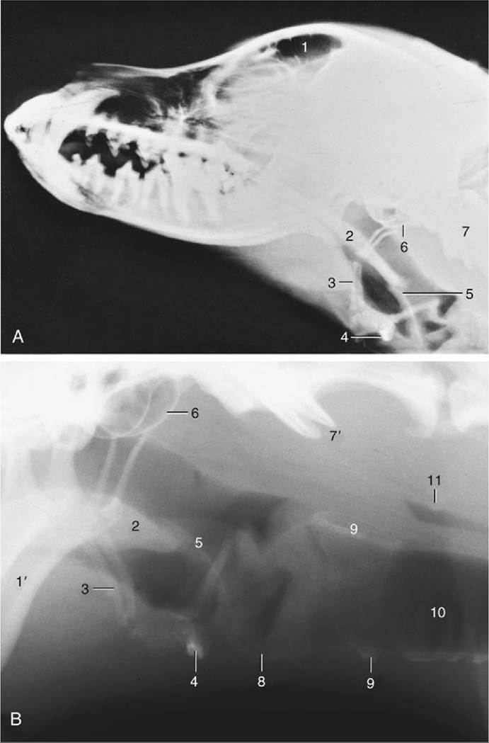
FIG. 11.32 (A) Radiograph of the canine head to show the relation of the hyoid apparatus to the skull and atlas. (B) Enlargement of the laryngeal region of another dog. 1, Frontal sinus, 1', mandible; 2, soft palate; 3, hyoid apparatus (epihyoid); 4, basihyoid; 5, epiglottis; 6, tympanic bulla; 7, atlas; 7', wings of atlas; 8, thyroid cartilage; 9, cricoid cartilage; 10, trachea; 11, air in esophagus.
Laryngeal paralysis as a genetic disorder occurs in certain breeds, notably the Bouvier and Leonberger, but it has also been encountered as an occasional disorder of older dogs of other large breeds.
The cranial laryngeal arteries provide the principal blood supply. They originate from the external carotid arteries and, with the cranial laryngeal nerves, pass through the rostral thyroid notches. Satellite veins drain into the external maxillary veins. Lymphatics drain into the medial retropharyngeal lymph nodes.
The cat's larynx is depicted in radiographs (see Fig. 11.29) and in a median section (see Fig. 11.35). The arytenoid cartilages have a simpler shape than those in the dog. The aryepiglottic folds bypass the arytenoid cartilages and connect the sides of the epiglottis directly to the cricoid cartilage. The vocal cords are thick and round; in contrast, the vestibular folds are thin and sharp edged. There is no genuine ventricle, but small pouches of the vestibular mucosa extend lateral to the fold. Solitary lymph nodules are present on the laryngeal surface of the epiglottis, and aggregated nodules (paraepiglottic tonsils) thicken the aryepiglottic folds.
Electromyographic studies show that purring in cats is produced by fast twitching of muscles in the larynx and diaphragm. The laryngeal muscles rapidly narrow and widen the glottis, causing respiratory air to vibrate and make the sound.
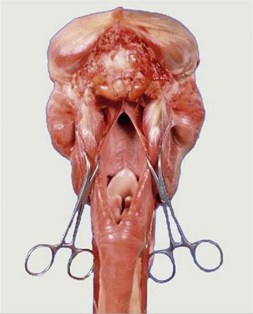
FIG. 11.33 The nasopharyngeal cavity exposed by median incision of the roof. Note the postvelar position of the tip of the epiglottis.
There are differences between the upper airways of brachycephalic and mesaticephalic breeds. In the brachycephalic obstruction syndrome, the nostrils can be stenotic, the pharynx short and narrow with thickened redundant mucosa, the root of the tongue massive, and the soft palate overlong. The progressive dyspnea is caused by increasing body weight, relatively insufficient growth of the laryngeal structures, increasing mass of the pharyngeal mucosa, and insufficient opening of the glottis. In addition, there is progressive collapse of the laryngeal structures and eversion of the laryngeal ventricles because of the increased traction caused by the greater velocity of exhaled air passing the relatively small laryngeal opening.