THE LARYNX, TRACHEA, AND SYRINX
The larynx occupies a mound on the floor of the oropharynx (see Figure 37-14/5). It is supported by cricoid and paired arytenoid cartilages that differ markedly from their mammalian counterparts but occupy similar positions.
The arytenoids articulate with the rostrodor- sal part of the annular cricoid. The glottis, formed by the arytenoids, closes the entrance to the larynx by reflex muscular action, preventing food particles and other foreign matter from reaching the lower air passages. Despite the narrowness of the glottis, it is possible to intubate the trachea in larger cage birds. There are no vocal folds; voice production occurs in the syrinx, a specialization at the tracheal bifurcation.The trachea, composed of tightly stacked, complete, and overlapping cartilaginous rings, accompanies the esophagus through the neck; it can be palpated on the right side (Figure 37-15). In a long-necked species, for example, trumpeter swans and cranes, it is much longer than the neck and forms a loop that is accommodated in an excavation of the sternum at the thoracic inlet. The trachea bifurcates into two primary bronchi dorsal to the base of the heart. These enter the ventral surface of the lungs after short course. In penguins, a median septum divides the trachea into left and right tubes, making it very easy to intubate a primary bronchus by mistake.
The syrinx is formed by the terminal part of the trachea and first parts of the primary bronchi (Figure 37-26). The tracheal cartilages of the syrinx are sturdy, but the bronchial cartilages are largely lacking, although a short vertical bar (pessulus; Figure 37-26/3) separates
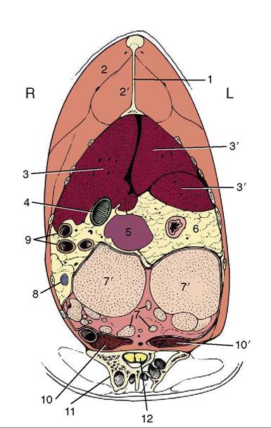
Figure 37-24 Transverse section of the trunk at the cranial end of the ilium. 1, Keel of sternum, 2, pectoralis; 2', supra- coracoideus; 3, 3', right and left lobes of liver; 4, gallbladder; 5, spleen; 6, constriction between proventriculus and gizzard; 7, ovary; 7’, follicle; 8, cranial mesenteric vein in mesenteric fat; 9, small intestine; 10,10', right and left kidneys; 11, ilium; 12, spinal cord.
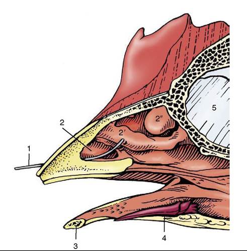
Figure 37-25 Section through rostral part of the head of a chicken.
1, Wire in nostril; 2, 2’, 2", rostral, middle, and caudal nasal conchae; 3, mandible; 4, tongue; 5, interorbital septum.the bronchial openings. The lateral and medial walls of the initial segments of the bronchi are membranous and produce the voice when caused to flutter (Figure 37-26Z2,2'). The male duck and swan have an osseous bulla (believed to be a resonator) on the left side of the syrinx. In psittacines a median pessulus is missing. A small paired muscle, the sternotrachealis (Figure 37-16Z 5), pulls the trachea toward the syrinx and aids in vocalization. An elaborate set of five pairs of syringeal muscles is present in Passeriformes (songbirds), and the surrounding interclavicular air sac gives the voice resonance by pushing against these membranes. Despite their great speaking ability, parrots have a relatively simple syringeal apparatus with only three pairs of syringeal muscles.
Because the trachea is narrowed at the syrinx, this is a common site of obstruction by seeds or other foreign bodies or by fungal granulomas. Birds exhibiting voice changes should have the syrinx examined endoscopically. Other common causes of voice changes are goiter
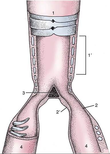
Figure 37-26 Semischematic representation of the opened syrinx. 1, Trachea; 1', tympanum; 2, 2’, lateral and medial tympaniform membranes; 3, pessulus; 4, primary bronchi.
pressing the syrinx or Aspergillus infection of the surrounding interclavicular air sac.