THE LIVER
The liver (hepar) is located in the most cranial part of the abdomen, immediately behind the diaphragm. It is by far the largest gland in the body and performs many functions essential for life.
The most obvious is the production of bile, but the parts it plays in protein, carbohydrate, and fat metabolism are even more important and depend on the liver’s situation astride the bloodstream draining the gastrointestinal tract. This ensures that the products of digestion, which are conveyed in the bloodstream after absorption, are presented to the hepatic cells before entering the general circulation.The metabolic functions of the liver explain the wide interspecific variation in size: average values are about 3% to 5% of body weight in carnivores, 2% to 3% in omnivores, and as little as 1% to 1.5% in herbivores. The
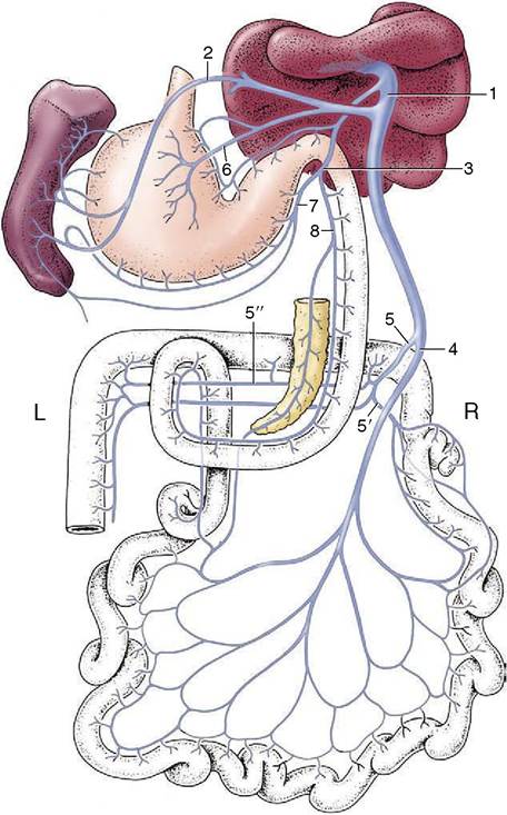
Figure 3-50 Semischematic dorsal view of the formation of the portal vein (dog). 1, Portal vein; 2, splenic vein; 3, gastroduodenal vein; 4, cranial mesenteric vein; 5, caudal mesenteric vein; 5, ileocolic vein; 5'l middle colic vein; 6, left gastric vein; 7, right gastroepiploic vein; 8, cranial pancreaticoduodenal vein.
liver is substantially heavier in the young animal than in the adult; it often shows considerable atrophy in old age. Usually brownish-red, the fresh liver is soft and has a characteristic friable consistency.
The adult liver intervenes between the diaphragm cranially and the stomach and intestinal mass caudally. Although extended across the median plane, the bulk lies to the right in all species (Figure 3-51). It is not so very asymmetrical in the dog: the proportions to the right and left of the median plane are about 3:2. In most species, including the dog, the liver is grossly divided into lobes by a series of fissures that extend inward from the ventral margin (Figure 3-52).
The lobation pattern shows many features of resemblance among different mammals, and considerable effort has been given to determining the homologies of individual lobes and fissures. The theoretical pattern, which accords the dog’s liver left lateral, left medial, right lateral, right medial, quadrate, and caudate lobes, of which the last is enlarged by papillary and caudate processes, is illustrated (Figure 3-53). It should not be regarded as more than a convenient fiction that facilitates description. Modern studies minimize the significance of the external fissuration and rely more on the internal ramifications of the vessels to establish homologies. Such studies have had the useful by-product of providing the surgeon with the detailed knowledge of the vascular architecture necessary for the safe removal of diseased parts of the human liver.In life the liver adapts to the form of neighboring organs, and when fixed in situ, it retains the conformation and impressions these impose. The rather large liver of the dog is therefore bluntly conical, and its cranial surface matches the curvature of the diaphragm against which it is pressed. The caudal surface is concave; to the left it exhibits a large excavation for the stomach, which is then extended over the median plane into a narrow duodenal groove. The dorsal border extends more cau- dally and reaches farther dorsally on the right side, where it is further extended by the caudate process, which carries a deep impression for the cranial pole of the right kidney. Toward the median plane, this border carries a groove for the passage of the caudal vena cava and, to the left of this, a notch for the esophagus. The gallbladder lies between the quadrate and right medial lobes; it is partly attached, partly free, and in some dogs so deeply embedded that it reaches the parietal surface, thus making contact with the diaphragm (see Figure 3-53).
The liver is clothed in peritoneum except for relatively small areas at the porta (hilus), in the fossa for the gallbladder, and at the origin of certain peritoneal reflections.
The right and left triangular, the coronary, and the falciform ligaments that pass to the diaphragm from the parietal surface have fibrous cores and attach the liver firmly; the lesser omentum, which passes from the visceral surface to the stomach and duodenum, is more fragile. A tunica fibrosa encloses the parenchyma beneath the serosa; it enters the substance at the porta and detaches extensions that convey the blood vessels inward, dividing where the vessels divide and thinning at each division. The finer trabeculae pervade the entire organ and divide the liver into innumerable small units, the hepatic lobules of the classic description. Although particularly marked in the pig’s liver (Figure 3-54), the lobular pattern is also quite obtrusive in that of the dog, in which the lobules appear as hexagonal areas (about 1 mm across) on the intact surface and in gross and histological sections.The liver receives a very generous blood supply through the hepatic artery, a branch of the celiac artery, and the portal vein. The relative importance of these
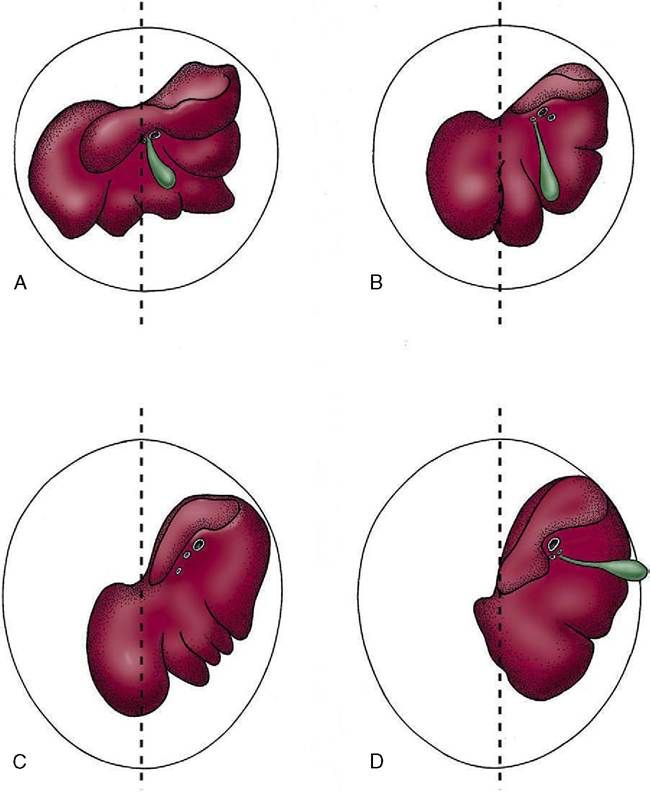
Figure 3-51 Caudal surface of the liver of the dog (A), pig (B), horse (C), and cattle (D). The median planes are indicated. The liver is asymmetrical, less so in the dog, more so in the pig and horse, and most in cattle, in which the bulk of the organ is displaced to the right. Note the absence of a gallbladder from the horse liver.
two supplies varies among species. The proportions are not known with certainty for the dog; the artery supplies the human liver with only one fifth of the blood but about three fifths of the oxygen. The branches of the hepatic artery that actually enter the liver are effectively end-arteries. However, provision exists for a collateral circulation outside the liver, between the hepatic artery and the other branches of the celiac artery that supply the stomach and duodenum (see Figure 3-39).
The intrahepatic arteries divide in company with branches of the portal vein and tributaries of the hepatic duct. They supply the connective tissue structures en route to the hepatic sinusoids into which both they and the branches of the portal vein eventually discharge.The portal vein is formed by the union of tributaries draining the digestive tract, pancreas, and spleen (see Figure 3-50). It is connected to systemic veins in the cardioesophageal and rectoanal regions at the extremities of its territory. These connections provide alternative outlets for portal blood when the flow through the liver is obstructed or impaired. The effects of obstruction vary between species and reflect the varying effectiveness of the hepatic artery in supplying oxygen. In the dog complete obstruction is rapidly fatal.
All blood delivered to the liver is collected by a single set of veins of which the central veins of the hepatic lobules are the smallest radicles. These eventually form the few large hepatic veins that open into the caudal vena cava as this tunnels through the liver substance. The circulation through the liver possesses numerous anastomoses—interarterial, intervenous, and arteriovenous; it is also controlled by various sphincter mecha-
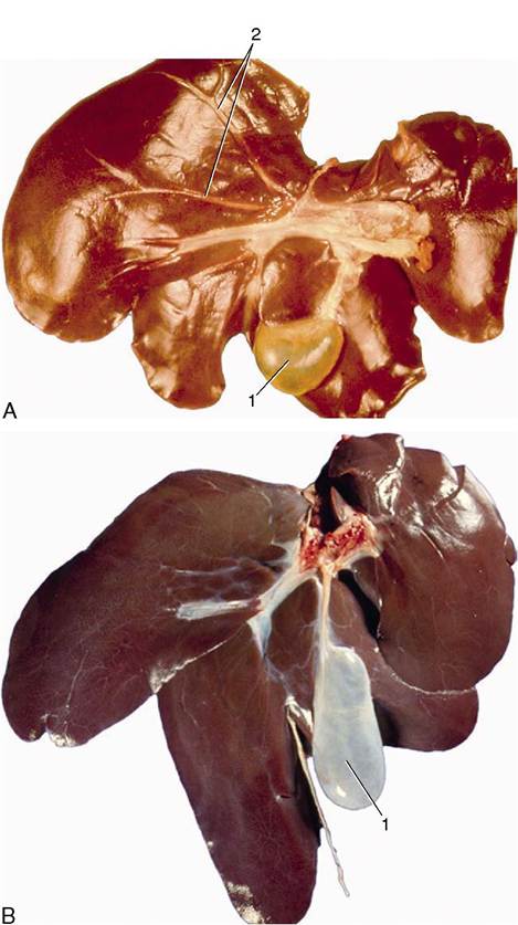
Figure 3-52 A, Visceral surface of liver (dog). B, Visceral surface of liver (pig). 1, Gallbladder; 2, hepatic ducts.
nisms, and together these features make it capable of very subtle regulation. A relatively rare congenital defect allows portal blood to pass directly to the caudal caval vein.
The liver receives sympathetic and parasympathetic nerves by way of periarterial plexuses and the vagal trunks, respectively.
The hepatic duct system begins with microscopic canaliculi within the lobules. These open into larger ductules that ultimately form a few large hepatic ducts by successive unions within the connective tissue between the lobules.
Before or shortly after leaving the liver at the porta these combine in a single trunk that runs to the duodenum (Figure 3-55). A tortuous side branch (cystic duct) that arises from the common trunk leads to the pear-shaped gallbladder. The part of the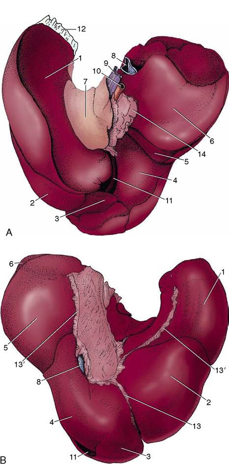
Figure 3-53 Visceral (A) and diaphragmatic (B) surfaces of the canine liver. 1, Left lateral lobe; 2, left medial lobe; 3, quadrate lobe; 4, right medial lobe; 5, right lateral lobe; 6, caudate process (of caudate lobe); 7, papillary process (of caudate lobe); 8, caudal vena cava; 9, portal vein; 10, hepatic artery; 11, gallbladder; 12, left triangular ligament; 13, falciform ligament; 13', coronary ligaments; 14, lesser omentum.
common trunk that is distal to the origin of the cystic duct is known as the bile duct (ductus choledochus). Variation in the duct system is frequent; some hepatic ducts may enter the gallbladder directly, while others may join the main outlet distal to the cystic duct. The gallbladder not only stores the bile but also concentrates it by absorption through the folded mucosa. As
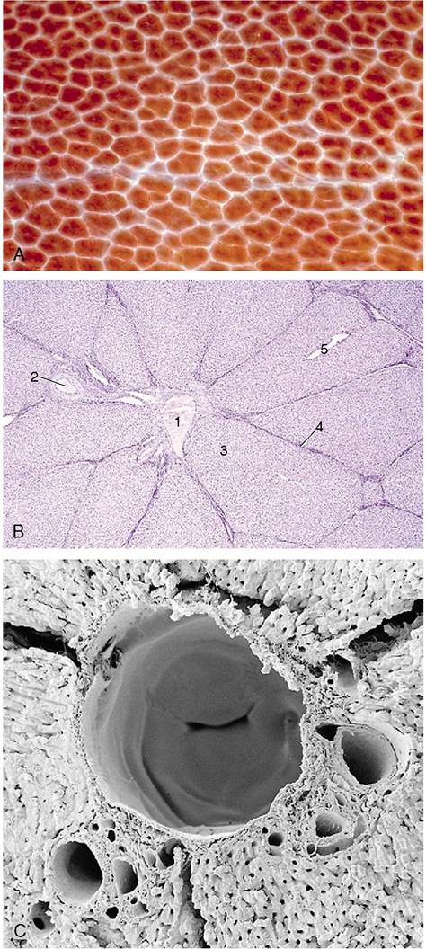
Figure 3-54 A, Surface of liver (enlarged) with clearly defined hepatic lobules (pig). B, Liver (pig) (28?). 1, central v.; 2, interlobular a.; 3, hepatic lobule; 4, interlobular connective tissue; 5, centrolobular venule. C, Scanning electron microscopy of corrosion cast of hepatic vessels (rat); note valve within central v.
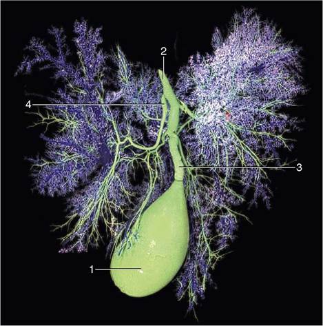
Figure 3-55 The bile drainage system of the dog. 1, Gallbladder; 2, bile duct; 3, cystic duct; 4, hepatic ducts.
is well known, a gallbladder is not essential; it is lacking in the horse, the rat, and certain other species, which compensate by enlargement of the duct system (see Figure 3-51).
The muscle of the bladder wall and duct, including the sphincter at the entrance to the duodenum, is supplied by parasympathetic nerves. Pain arising from the duct system, common in human patients, is abolished by section of the (sympathetic) splanchnic nerves.