THE LIVER
The liver is quite variable in form and size but on average weighs about 5 kg in a saddle horse, thus accounting for about 1.5% of the body weight, a much smaller proportion than in carnivores.
It is situated in the most cranial part of the abdomen directly against the diaphragm. It is markedly asymmetrical in the healthy young subject, in which about two thirds lie to the right of the median plane (see Figure 21-7/2). The most caudal part, which is also the most dorsal, lies ventral to the vertebral extremities of the sixteenth and seventeenth ribs of the right side; the most cranial and most ventral part lies against the left part of the vertex of the diaphragm (Figure 21-6/5). The long axis thus runs obliquely. In the newborn foal the liver is relatively much larger and extends onto the abdominal floor behind the costal arch; it is also more symmetrical. In older subjects atrophy is common; this is most obvious in the right lobe and probably results from chronic pressure from the right dorsal colon and cecal base. Less often, the left lobe atrophies, perhaps under pressure from the stomach.
The parietal surface is joined to the diaphragm by a complicated system of ligaments. The visceral surface lies against and is impressed by the stomach, duode-
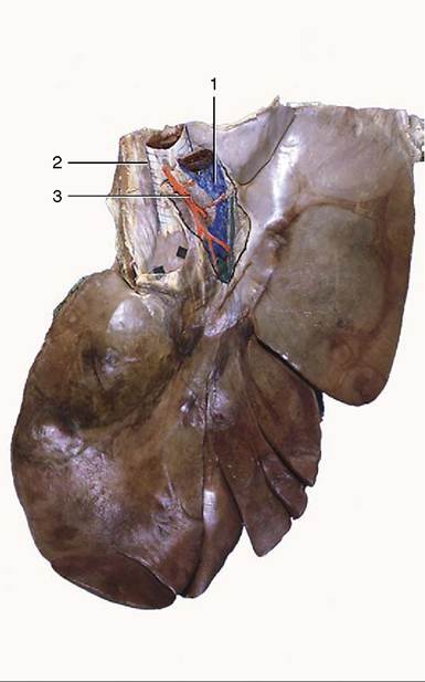
Figure 21-20 Visceral surface of the liver. 1, Portal v.;
2, caudal vena cava; 3, hepatic a.
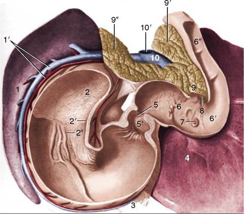
Figure 21-21 Topography of spleen, stomach, pancreas, and liver, caudoventral view. 1, Intestinal surface of spleen; 1', splenic a. and v.; 2, fundus (blind sac) of stomach; 2', cardia; 2", margo plicatus; 3, greater omentum; 4, liver; 5, pyloric orifice; 5', pyloric antrum; 6, S-shaped cranial part of duodenum; 6,, cranial flexure of duodenum; 6", descending duodenum; 7, major duodenal papilla; 8, minor duodenal papilla; 9, body of pancreas; 9' (right)—9" (left), left and right lobes of pancreas; 10, portal v; 10', stump of cranial mesenteric v.
num, dorsal diaphragmatic flexure of the colon, and cecal base (see Figure 21-10).
The porta is central, within an area made rough by the direct attachment of the pancreas. The dorsal fixed margin of the liver extends between the right and left triangular ligaments and is very irregular (Figure 21-20). Its right part is thick and excavated to receive the cranial pole of the right kidney; a sulcus medial to this transmits the caudal vena cava. Its left part is much thinner and does not extend nearly so far dorsally; it carries the impression of the esophagus close to the midline. The long free margin is much sharper and is interrupted by a series of fissures, of which the largest divide named lobes. The current nomenclature recognizes left, quadrate, right, and caudate lobes. The first two are separated by the fissure carrying the round ligament of the liver (vestige of the umbilical vein), but the boundaries of the others are more arbitrary and are of doubtful morphological significance.The duct system is remarkable for the absence of a gallbladder, but its wide caliber compensates for this. The bile duct opens into the cranial duodenum on the papilla shared with the major pancreatic duct (Figure 21-21/7). The oblique passage of the duct through the duodenal wall serves as a sphincter and prevents the influx of ingesta.