The Liver and Pancreas
The liver appears as an endodermal diverticulum at the junction of the foregut and midgut. It quickly divides into a cranial branch, which forms the gland tissue and hepatic ducts, and a caudal branch, which forms the gallbladder and cystic duct (Fig.
3.62).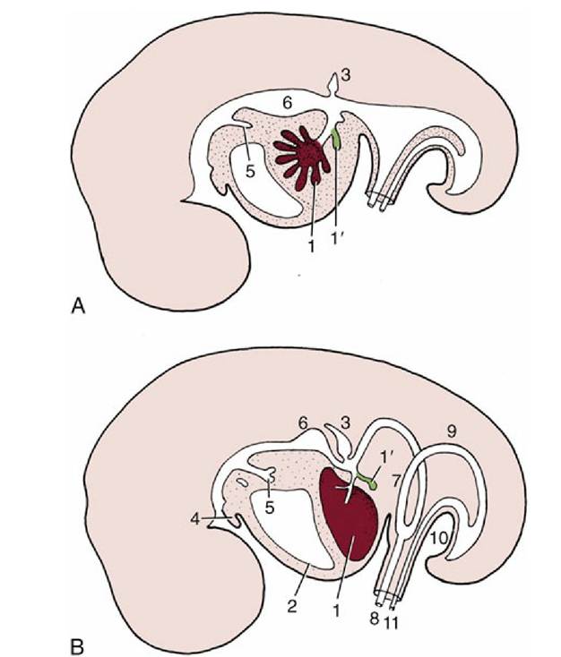
FIG. 3.62 Development of the liver. (A) Early development: a cranial branch (1) of the endodermal diverticulum invades the septum transversum; a caudal branch (1) forms the gallbladder and cystic duct.
(B) A later stage, in which the developing liver expands caudally into the abdominal cavity. 1, liver; 1', gallbladder; 2, pericardium and heart; 3, dorsal primordium of pancreas; 4, tongue; 5, tracheobronchial diverticulum; 6, stomach; 7, loop of midgut; 8, vitelline duct; 9, hindgut; 10, cloacal membrane; 11, allantoic stalk.
The cranial branch extends finger-like processes into the splanchnic mesoderm of the adjacent septum transversum, carried here with the formation of the head fold. As the processes penetrate the mesoderm, they engage with the vitelloumbilical system of veins, which arrive here from the extraembryonic membranes. Very soon a three-dimensional spongework of hepatic cell-cords and plates is formed, surrounded on all sides by thin-walled blood vessels, which is a precocious realization of the adult arrangement. Attenuation of the connection between the liver and the gut forms the lesser omentum.
The growth of the liver, extremely rapid in younger embryos, is a major factor in the temporary herniation of the midgut (see later). Although its growth slows later, the liver remains disproportionately large (in comparison with that in the adult) until well after birth. One relevant factor is the exercise of an erythropoietic activity before birth that is later relinquished.
The secretory and metabolic functions are established by midterm in the human fetus.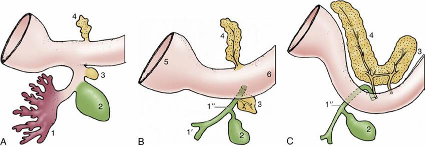
FIG. 3.63 Development of the pancreas. (A) Early stage. (B) A later stage showing separate duct systems in the two primordia. (C) The two primordia have fused after the migration of the ventral pancreas. The dorsal pancreas now drains mainly via the ventral duct system. 1, Liver primordium; 1', hepatic ducts; 1", bile duct; 2, gallbladder; 3, ventral primordium of pancreas; 4, dorsal primordium of pancreas; 5, stomach; 6, duodenum.
The pancreas arises from the same portion of the foregut as the liver. There are initially two primordia: one is dorsal and the second is ventral and associated with the hepatic outgrowth (Fig. 3.63). These later fuse, allowing combination of the two duct systems, following which one or the other may lose its connection with the gut. The islet tissue develops by budding from the ducts. Both endocrine and exocrine components are competent well before birth.
The celiac artery is associated with the postpharyngeal part of the foregut.