THE LYMPHATIC STRUCTURES OF THE THORAX
The thoracic lymph nodes, arranged in four centers (Figure 33-3/6 and Figure 33-4/7), collect lymph from
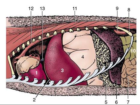
Figure 33-4 The heart in situ.
1, Heart; 2, diaphragm; 3, left lobe of liver; 4,stomach, greatly dilated; 5,greater omentum, gastrosplenic ligament; 6, spleen; 7, jejunum; 8, last rib; 9, left kidney; 10, ascending colon; 11, back muscles; 12, aorta; 13, caudal vena cava.the thoracic walls and contents and from adjacent structures and channel it to the thoracic duct or, where some more cranial nodes are concerned, directly into veins at the thoracic inlet.
The dorsal thoracic center comprises a variable number of small aortic nodes that receive lymph from the dorsal part of the thoracic wall, the mediastinum, and mediastinal nodes. The ventral center consists of fewer but larger sternal nodes concerned with the ventral part of the thoracic walls and the first two or three pairs of mammary glands.
Inconstant numbers of cranial and caudal mediastinal nodes form a chain above the base of the heart. The cranial nodes drain structures of the neck in addition to mediastinal contents, including the tracheobronchial nodes. Their efferents are divided into some that open into veins directly and others that lead to the thoracic duct. The caudal nodes are not always to be found. When present, they drain neighboring structures and send their efferents to the tracheobronchial and aortic nodes.
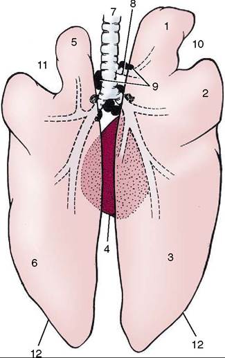
Figure 33-5 The lungs, dorsal view (see also Figure 4-23). 1, Right cranial lobe; 2, right middle lobe; 3, right caudal lobe; 4, accessory lobe of right lung; 5, divided left cranial lobe; 6, left caudal lobe; 7, trachea; 8, tracheal bronchus; 9, tracheobronchial lymph nodes; 10, right cardiac notch; 11, left cardiac notch; 12, basal border.
The bronchial center (Figure 33—3/4) consists of a dozen or so tracheobronchial nodes arranged about the origin of the bronchi (Figure 33-5). They drain the lungs, heart, and pericardium and in turn drain to the cranial mediastinal nodes or directly into the thoracic duct.
The thoracic duct runs from caudal to cranial between the aorta and esophagus, passing the trachea at its left side before joining the bloodstream.