THE THORAX
The body of a pig does not widen appreciably where the neck joins the trunk: the subcutaneous layer of fat allows the forelimb to blend in unobtrusively, and only a slight depression between the flabby jowls and the shoulder joint marks the junction.
There is a similar depression between the elbow joint and the thoracic wall. The “points” of both joints are palpable. The olecranon of the elbow projects onto the ventral end of the fifth rib (Figure 33-2). The manubrium of the sternum is also easily found.Most pigs have 14 or 15 pairs of ribs; asymmetry of number is common (see Figure 32-1). The first seven pairs are sternal. The rib cage is smaller than the external dimensions suggest; it is especially narrow and shallow between the forelimbs but deepens caudally with the upward sweep of the thoracic vertebrae. It is relatively long, depending to some extent on the number of vertebrae. The line of pleural reflection follows the dorsal half of the last rib before descending in a gentle curve to cross the seventh costochondral joint (Figure 33-2). The cranial mediastinum, like that of ruminants,
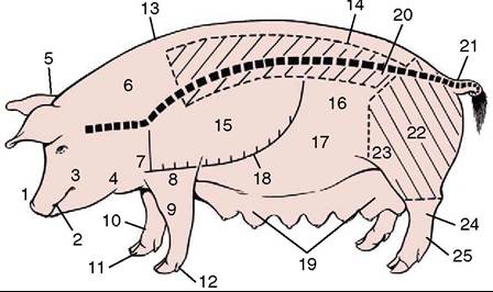
Figure 33-1 Parts of the pig. The position of the vertebral column is indicated. The hatched areas show ham and loin of the meat trade. 1, Snout; 2, mouth; 3, cheek; 4, jowls; 5, poll; 6, neck; 7, shoulder joint; 8, elbow joint; 9, carpus; 10, fetlock joint; 11, hoof; 12, accessory digit; 13, withers; 14, loin (lumbar area); 15, thorax; 16, flank; 17, abdomen; 18, ventral extent of bony thorax; 19, mammary glands; 20, position of coxal tuber; 21, tailhead; 22, thigh; 23, stifle joint; 24, hock joint; 25, metatarsus.
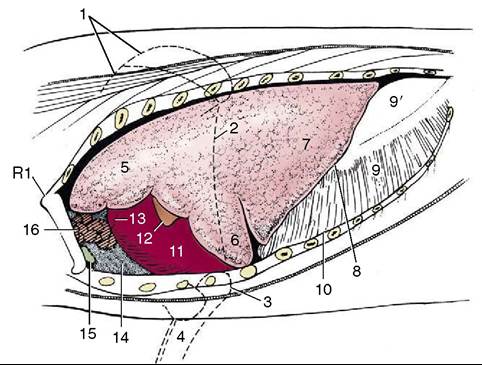
Figure 33-2 The thoracic viscera in situ, semischematic.
1, Scapula; 2, caudal border of triceps; 3, olecranon; 4, radius and ulna; 5, 6, cranial and caudal parts of cranial lobe of lung; 7, caudal lobe of lung; 8, basal border of lung; 9, 9', muscular and tendinous parts of diaphragm; 10, line of pleural reflection; 11, heart; 12, 13, left and right auricles; 14, cranial mediastinum; 15, sternal lymph node; 16, thymus.attaches to the ventral parts of the left first and second ribs, but more dorsally it is separated from the thoracic wall by the cranial lobe of the left lung.
The left lung possesses a cranial lobe, divided by a cardiac notch, and a caudal lobe (Figure 33-2/5,6,7 and see Figure 4-23). The right lung possesses cranial, middle, caudal, and accessory lobes, separating the cardiac notch separates the first two (see Figure 4-23, A). The cranial lobe of this lung is ventilated by a sepa-
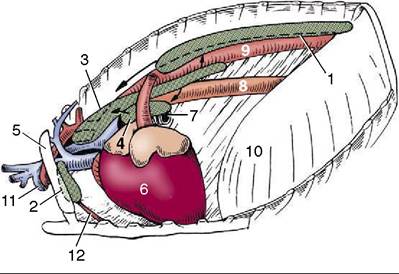
Figure 33-3 The lymph centers of the thorax, left lateral view. 1, Dorsal thoracic lymph center; 2, ventral thoracic lymph center; 3, mediastinal lymph center; 4, tracheobronchial lymph center; 5, first rib; 6, heart; 7, left bronchus; 8, esophagus; 9, aorta; 10, diaphragm; 11, axillary vein and artery; 12, internal thoracic artery.
rate tracheal bronchus (Figure 33-5/8 and see Figure 4-24). The lobulation of the lungs is relatively distinct.
The projection of the lungs onto the thoracic wall is small. The basal border of the left lung extends from the sixth costochondral junction to the upper end of the third last rib. This border of the right lung is less steep and reaches the penultimate rib. Auscultation and percussion of the lungs are usually reserved for young pigs of cooperative disposition.
The heart is small, providing as little as 0.3% of body weight (compared with 1.5% or more in athletic species such as the horse and dog), and this has been cited as a predisposing factor in “sudden death syndrome” commonly occurring in pigs. Heart size has not kept pace with the much-accelerated growth of modern, improved pigs, which reach a weight of 115 kg at 5 or 6 months; in striking contrast, 2 or 3 years was required to reach the much more modest weight of 40 kg in 1800. The heart occupies the ventral half of the thoracic cavity, extending between the second and fifth ribs (Figure 33-3/6 and Figure 33-4/7). It is thus covered by the forelimb in the standing animal but can be made accessible by drawing the limb forward. It exhibits no structural distinctions of note (see Figure 7-7).
Paracentesis is best performed through the fifth left or the fourth right intercostal space; the needle is inserted about 5 cm dorsal to the olecranon (Figure 33-2).