» The Lymphatic Structures of the Thorax
The thoracic lymph nodes, arranged in four centers (Fig. 33.8/1-4), collect lymph from the thoracic walls and contents and from adjacent structures and channel it to the thoracic duct or, where some more cranial nodes are concerned, directly into veins at the thoracic inlet.
The dorsal thoracic center comprises a variable number of small aortic nodes that receive lymph from the dorsal part of the thoracic wall, the mediastinum, and mediastinal nodes. The ventral center consists of fewer but larger sternal nodes concerned with the ventral part of the thoracic walls and the first two or three pairs of mammary glands.
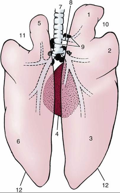
FIG. 33.4 The lungs, dorsal view (see also Fig. 4.23). 1, Right cranial lobe; 2, right middle lobe; 3, right caudal lobe; 4, accessory lobe of right lung; 5, divided left cranial lobe; 6, left caudal lobe; 7, trachea; 8, tracheal bronchus; 9, tracheobronchial lymph nodes; 10, right cardiac notch; 11, left cardiac notch; 12, basal border.
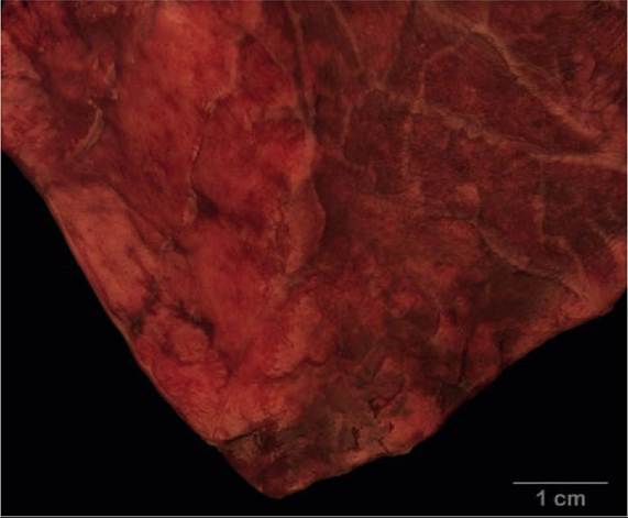
FIG. 33.5 Surface of pig lung showing lobulation.
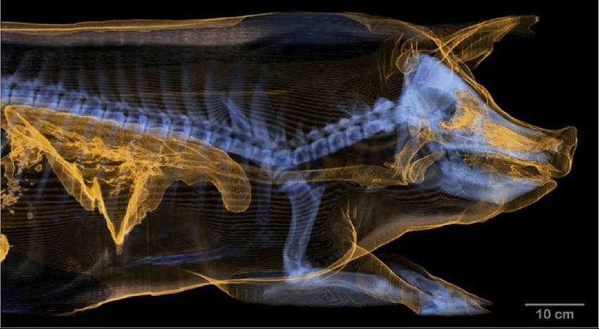
FIG. 33.6 Three dimensional projection generated from computed-tomographic imaging of gilt showing location of respiratory tract and lungs. Air in the nasal cavity, ethmoturbinates, larynx, trachea and lungs is shown in yellow. Skeleton is shown in blue.
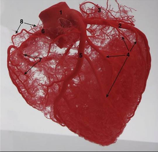
FIG. 33.7 Corrosion cast of the pig heart (auricular surface). 1, Aorta; 2, circumflex branch of the left coronary artery, also called the left circumflex artery (LCX); 3, atrial branches of the LCX; 4, ventricular branches for the left ventricle; 5, paraconal interventricular branch of the left coronary artery, also called the left anterior descending branch (LAD); 6, right coronary artery (RCA); 7, ventricular branches for the right ventricle.
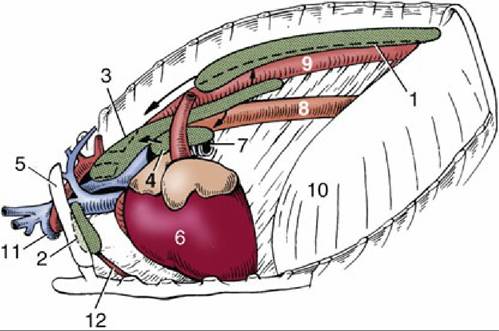
FIG.
33.8 The lymph centers of the thorax, left lateral view. 1, Dorsal thoracic lymph center; 2, ventral thoracic lymph center; 3, mediastinal lymph center; 4, tracheobronchial lymph center; 5, first rib; 6, heart; 7, left bronchus; 8, esophagus; 9, aorta; 10, diaphragm; 11, axillary vein and artery; 12, internal thoracic artery.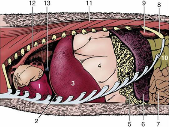
FIG. 33.9 The heart in situ, lateral view of dissected thorax. 1, Heart; 2, diaphragm; 3, left lobe of liver; 4, stomach, greatly dilated; 5, greater omentum, gastrosplenic ligament; 6, spleen; 7, jejunum; 8, last rib; 9, left kidney; 10, ascending colon; 11, back muscles; 12, aorta; 13, caudal vena cava.
Inconstant numbers of cranial and caudal mediastinal nodes form a chain above the base of the heart (Mediastinal lymph centre). The cranial nodes drain structures of the neck in addition to mediastinal contents, including the tracheobronchial nodes. Their efferents are divided into some that open into veins directly and others that lead to the thoracic duct. The caudal nodes are not always to be found. When present, they drain neighboring structures and send their efferents to the tracheobronchial and aortic nodes.
The tracheobronchial centre (Fig. 33.8/4) consists of a dozen or so tracheobronchial nodes arranged about the origin of the bronchi (Fig. 33.4/9). They drain the lungs, heart, and pericardium and in turn drain to the cranial mediastinal nodes or directly into the thoracic duct.
The thoracic duct runs from caudal to cranial between the aorta and esophagus, passing the trachea at its left side before joining the bloodstream.
Comprehension Check
Indicate the sites for paracentesis of the thorax.
Describe the major lymph collection centers in the thorax, and list the areas/organs serviced by them.