THE LYMPHATIC STRUCTURES OF THE HEAD AND NECK
Five lymph centers are located in the head and ventrolateral part of the neck (Figure 32-13). The mandibular center comprises about six principal and four accessory nodes. The mandibular nodes lie behind the caudoven- tral border of the mandible, related to the mandibular gland and crossed laterally by the facial vein (Figure
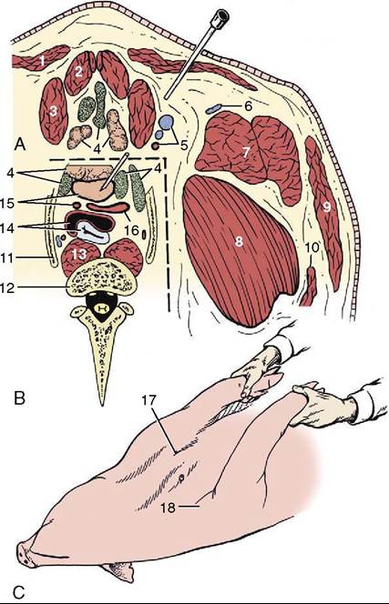
Figure 32-12 A, Transverse section of the ventral neck slightly cranial to the manubrium sterni.
B, The area within the broken line represents the topography at the slightly more caudal level of the first ribs. C, Pig held on its back for cranial vena cava venipuncture; see needle in position. 1, Cutaneous colli; 2, sternohyoideus; 3, sternocephalicus; 4, lymph nodes and thymus; 5, common carotid artery and external and internal jugular veins; 6, cephalic vein; 7, brachiocephalicus; 8, subclavius; 9, platysma; 10, omotransversarius; 11, first rib; 12, body of C7; 13, longus colli; 14, trachea and esophagus; 15, cranial vena cava and left subclavian artery; 16, bicarotid trunk and right subclavian artery; 17, palpable manubrium sterni; 18, shoulder joint.32-14/7). They drain the ventral half of the head and forward lymph to the accessory group and to ventral and dorsal superficial cervical nodes and are routinely examined in meat inspection. The accessory nodes (Figure 32-14/2) are also located by the border of the mandible and under cover of the parotid gland. They drain the same part of the head and also the ventral part of the neck; their efferents also go to the superficial cervical nodes. The parotid nodes (Figure 32-14/3) are
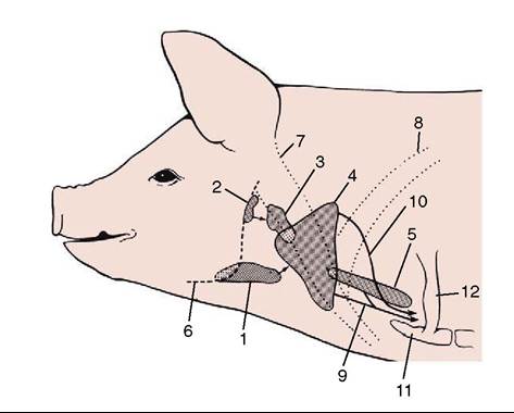
Figure 32-13 The lymph centers of the head and neck, schematic. The arrows indicate lymph flow.
1, Mandibular lymph center; 2, parotid lymph center; 3, retropharyngeal lymph center; 4, superficial cervical lymph center; 5, deep cervical lymph center; 6, mandible; 7, brachiocephalicus; 8, subclavius; 9, tracheal lymph trunk; 10, lymph from dorsal superficial cervical nodes; 11, manubrium sterni; 12, first rib.located ventral to the temporomandibular joint covered by the parotid gland. They drain the head dorsal to the palate and send their efferents to the lateral retropharyngeal nodes (Figure 32-14∕√).
The retropharyngeal center consists of one medial and two lateral nodes (Figure 32-14∕√,5). The latter lie near the joint, again under the parotid gland and a few centimeters caudal to the parotid center. They drain superficial structures where the head joins the neck; their efferents go to the dorsal superficial cervical nodes. The medial node lies above the pharynx and drains deeper structures at the same level as the lateral nodes; its efferents join to form a tracheal duct.
The superficial cervical center consists of about 10 nodes, roughly arranged in a triangle and divided into dorsal, middle, and ventral groups (Figure 32-14/0). Together, they correspond to the single group found deep to the omotransversarius in other species. The dorsal nodes drain the neck and neighboring parts of the thoracic wall and forelimb. They also receive lymph from the head nodes, other than the medial retropharyngeal, and pass it to veins at the thoracic inlet. The middle group is dorsal to the external jugular vein and drains the shoulder region; its efferents accompany or join those of the dorsal group. The ventral group is arranged in a chain and, like the middle nodes, lies deep to the brachiocephalic muscle. It drains superficial
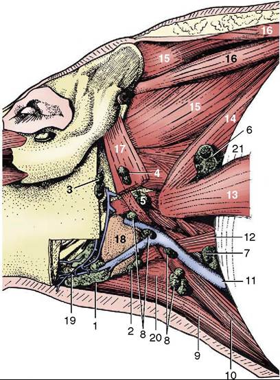
Figure 32-14 Dissection of the neck to show the lymph nodes, left lateral view. 1, Mandibular lymph nodes; 2, accessory mandibular lymph nodes; 3, parotid lymph nodes; 4, lateral retropharyngeal lymph nodes; 5, medial retropharyngeal lymph nodes; 6-8, dorsal, middle, and ventral superficial cervical lymph nodes; 9, sternohyoideus; 10, sternocephalicus; 11, external jugular vein; 12, omohyoideus; 13, omotransver- sarius; 14, serratus ventralis cervicis; 15, splenius; 16, rhomboideus cervicis et capitis; 17, cleidomastoideus; 18, mandibular gland; 19, facial vein; 20, thyrohyoideus; 21, subclavius.
structures of the neck, the forelimb, the ventral thoracic wall, and the first two mammary glands. It also receives lymph from the mandibular and lateral pharyngeal nodes.
In theory, the many nodes of the deep cervical center are divided into several groups spread at intervals along the internal jugular vein. In practice, few are usually to be found. They drain directly to the large veins at the thoracic inlet.