THE VENTRAL ASPECT OF THE NECK
The visceral space of the neck has the same contents as in other species and is similarly enclosed ventrolaterally by a series of thin, straplike muscles. The cutaneous muscle is thick at its origin from the manubrium but thins when followed cranially to merge with the cutane-
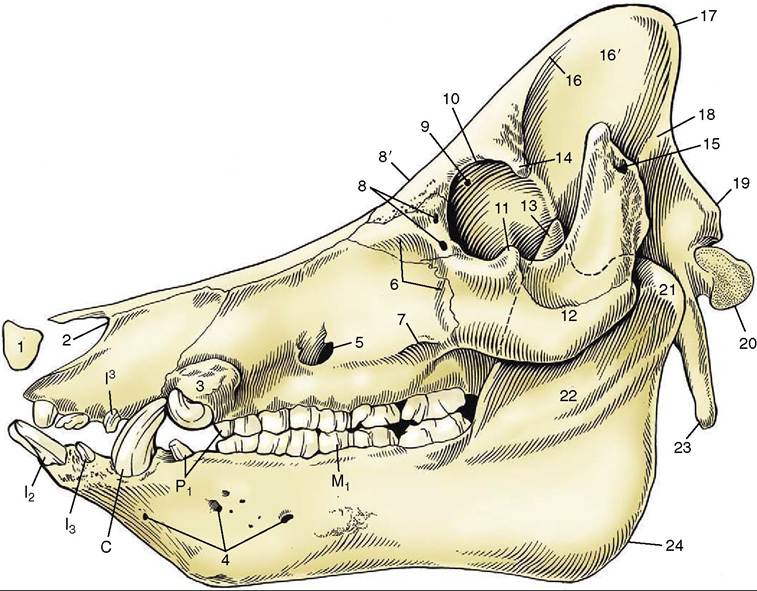
Figure 32-8 Skull of a boar.
1, Rostral bone; 2, nasoincisive notch; 3, canine eminence; 4, lateral mental foramina; 5, infraorbital foramen; 6, fossa canina; 7, facial crest; 8, lacrimal foramina; 8’, location of supraorbital foramen on dorsal surface; 9, orbital end of supraorbital canal; 10, orbital rim; 11, frontal process of zygomatic bone; 12, zygomatic arch; 13, coronoid process of mandible; 14, zygomatic process of frontal bone; 15, external acoustic meatus; 16, temporal line; 16', temporal fossa; 17, nuchal crest; 18, temporal crest; 19, nuchal tubercle; 20, occipital condyle; 21, condylar process of mandible; 22, ramus of mandible; 23, paracondylar process; 24, angle of mandible; I2,13,13, incisors; C, canine teeth (tusks); P1, first premolars; M1, first molar.
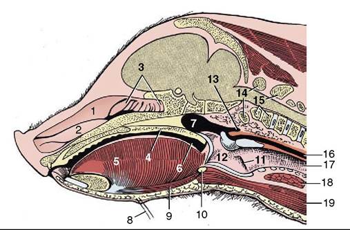
Figure 32-9 Median section of the head of a 4-week- old pig; the nasal septum has been removed. 1, Dorsal nasal concha; 2, ventral nasal concha; 3, ethmoidal conchae; 4, soft palate; 5, tongue; 6, oropharynx; 7, nasopharynx; 8, mental hairs; 9, geniohyoideus; 10, basihyoid; 11, laryngeal ventricle; 12, larynx; 13, pharyngeal diverticulum; 14, atlas; 15, axis; 16, esophagus; 17, trachea; 18, thyroid gland; 19, sternohyoideus.
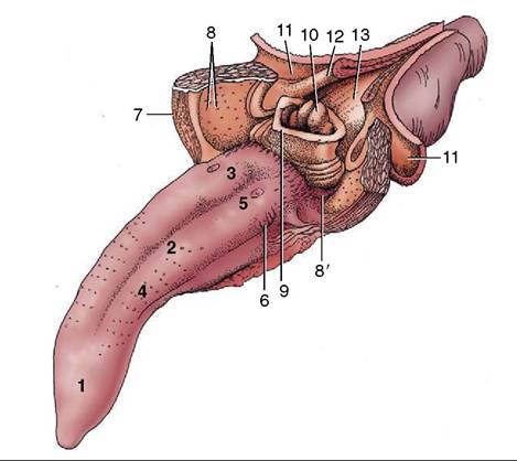
Figure 32-10 Tongue and pharynx.
The soft palate and the dorsal wall of the esophagus have been split in the midline. 1-3, Apex, body, and root of tongue; 4, fungiform papillae; 5, vallate papillae; 6, foliate papillae; 7, palatoglossal arch; 8, tonsil of the soft palate; 8', paraepiglottic tonsil; 9, epiglottis; 10, corniculate processes of the arytenoid cartilages; 11, dorsal wall of nasopharynx; 12, palatopharyngeal arch; 13, entrance to esophagus.ous muscles of the face. A more important impediment to puncture of the external jugular vein is the thick subcutaneous fat.
The trachea and esophagus show no unusual features nor do the vessels and nerves passing between head and thorax, apart from the internal jugular vein, which is considerably better developed than in most other species. The thyroid gland consists of two lobes, broadly connected ventral to the trachea; because of the shortness of the neck, it lies close to the thoracic inlet (see Figure 6-4, D). The thymus lies to each side of the larynx and trachea (Figure 32-11/3,4) and is particularly well developed: it does not attain its greatest size until the animal is about 9 months old and begins to regress a few months later. Its bulbous cranial extremity carries on its surface the minute (1 to 4 mm) external parathyroid glands. (The internal parathyroid glands are thought to disappear in the embryo.)
The most common clinical procedure involving the neck is cranial vena cava puncture, which may be performed in the standing animal or in one suitably restrained on its back. The needle is inserted in the depression between the manubrium and the point of the right shoulder and advanced in the direction of the left
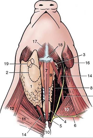
Figure 32-11 Ventral view of the neck. Deep dissection to the right; superficial dissection, from which the cutaneous colli has been removed, to the left; semischematic. 1, Parotid gland; 2, mandibular gland; 3, thymus—dot on cranial end indicates the position of the external parathyroid; 4, thyroid; 5, external jugular vein; 6, cephalic vein; 7, sternohyoideus (drawn narrower than actual width); 8, internal jugular vein; 9, larynx; 10, manubrium sterni; 11, superficial pectoral muscle; 12, brachiocephalicus; 13, subclavius; 14, sterno- cephalicus; 15, omohyoideus; 16, angle of mandible; 17, mylohyoideus; 18, basihyoid; 19, mandibular lymph nodes.
scapula until it meets one or other of the large veins between or just in front of the first pair of ribs. Entry is best made from the right because the left phrenic nerve is more vulnerable to injury; the thoracic duct also lies more to that side (Figure 32-12).