THE MAJOR NERVES OF THE HINDLIMB
It is only necessary to deal briefly with the course, relations, and distribution of those nerves that extend substantially into the free limb because a general account of the lumbosacral plexus (usually formed by the nerves L4-S2) and its divisions has been presented (pp.
323-325).The femoral nerve (L4-L6) has a very short course within the thigh before it ends by ramifying within the quadriceps femoris, the principal extensor of the stifle and an ancillary flexor of the hip. Shortly before disappearing into this muscle, it detaches the saphenous nerve, which descends subcutaneously over the medial aspect of the limb accompanied by the palpable saphe-
1
3
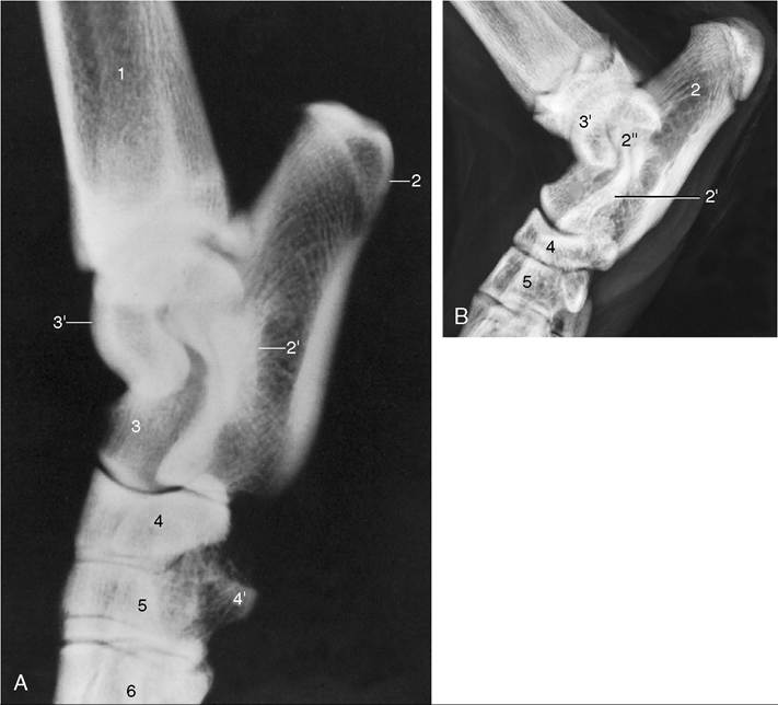
Figure 17-8 Lateral radiographic views of the canine (A) and young feline (B) hocks. 1, Tibia and fibula; 2, calcaneus; 2', sustentaculum tali; 2", coracoid process; 3, talus; 3', trochlea of talus; 4, superimposed fourth and central tarsal bones; 4', plantar tubercle on fourth tarsal bone; 5, distal row of tarsal bones; 6, metatarsal bones.
nous artery. Although the saphenous nerve supplies the sartorius, it is largely sensory, serving the skin of the medial surface of the thigh, stifle, leg, and hock (Figure 17-9). Dysfunction of the femoral nerve paralyzes the quadriceps, resulting in the collapse of the stifle and disabling the entire limb. Compensation is not available. The skin of the medial surface of the limb is deprived of sensation.
The sciatic nerve (L6-S1) crosses the dorsal border of the hip bone to enter the limb together with the caudal gluteal vessels. After passing the dorsocaudal aspect of the hip joint deep to the greater trochanter, where it is susceptible to injury in trauma or surgery of the joint, the nerve and accompanying vessels supply branches to the hamstring muscles.
The nerve then continues distally in a central position within the thigh, caudal to the femur and cushioned between the biceps laterally, the adductor, and later the semimembranosus medially (Figure 17-2/4). At a rather variable point it divides into common peroneal and tibial nerves that continue the course of the parent trunk until they diverge caudal to the stifle. The sciatic nerve and its peroneal and tibial branches collectively supply the skin of the entire limb distal to the stifle with the exception of the medial strip claimed by the saphenous.The common peroneal nerve, the more lateral of the terminal divisions of the sciatic, can be palpated in lean dogs where it passes over the lateral head of the gastrocnemius (Figure 17-5/7). It then dives deeply among the dorsal crural muscles (the extensors of the digits and flexors of the hock), which it supplies. It is continued by superficial and deep (peroneal) branches that enter the paw over the dorsal aspect of the hock; they supply the skin of the dorsal surface. Paralysis of the common peroneal nerve produces slight overextension of the hock and inability to extend the digits, which may be rested on their dorsal surfaces. In time, affected dogs learn to flick their paws forward before putting them down, enabling their limbs to support weight. The dorsal surface of the paw is without sensation.
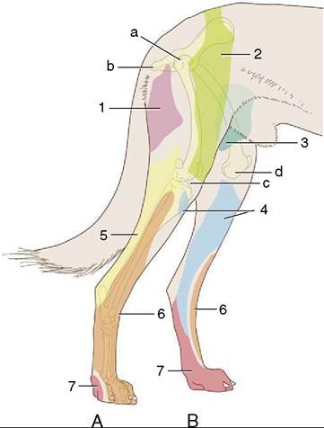
Figure 17-9 Autonomous zones of the cutaneous innervation on the lateral (A) and medial (B) surfaces of the canine hindlimb. 1, Caudal cutaneous femoral nerve (purple); 2, lateral cutaneous femoral nerve (green); 3, genitofemoral nerve (teal); 4, saphenous nerve (blue); 5, sciatic nerve (yellow); 6, peroneal nerve (orange); 7, tibial nerve (red). a, Position of greater trochanter; b, ischial tuber; c, lateral tibial condyle; d, medial tibial condyle.
The tibial nerve passes between the two heads of the gastrocnemius, where it detaches branches to the muscles behind the tibia (the flexors of the digits and extensors of the hock). The depleted nerve, now largely sensory but retaining a small motor component for the intrinsic muscles of the paw, continues distally within the web of skin between the caudal crural muscles and the common calcanean tendon. It crosses the hock beside the deep flexor tendon before branching to supply the plantar structures of the paw. Tibial nerve injuries cause the hock to be flexed and lowered closer to the ground when the limb bears weight. The paralysis of the digital flexors elevates the toes; their plantar aspect is without sensation.