CONFORMATION AND EXTERNAL FEATURES
The general character of the head is determined by the age, the sex, and the breed. In young foals the cranial vault is domed to match the contours of the brain and projects above a face that is both short and shallow (Figure 18-1).
The adult conformation develops as the face lengthens and deepens to accommodate the full complement of teeth and expanding paranasal sinuses; enlargement of the frontal sinus smoothes the dorsal profile at the junction of the face and cranium. Sex and breed differences are not wholly separable from those due to age, as the face is disproportionately increased in larger animals; a longer face is therefore characteristic of the adult compared with the juvenile, the stallion with the mare, and the heavy draft horse with the pony. The other very obvious breed difference concerns the dorsal profile; a relatively straight profile is generally preferred but some convexity (“ram’s head”) is characteristic of certain heavy breeds, whereas concavity (“dishing”) is the rule in Arabs and common in horses with admixture of Arab blood (see Figure 18-1). The ventral margin of the lower jaw of young horses may be disfigured by one or more rounded swellings, each corresponding in position to the root of an unerupted permanent cheek tooth. The temporary irregularities, though unsightly, are part of a normal process (p. 514).The skin is thinner and more firmly bound down than over most other parts of the body and is especially tight where it lies directly on bone. The coat is generally short, but a forelock continuing the mane may be prominent; a “mustache” is a feature of some animals, especially the larger breeds. Tactile hairs are numerous and widely scattered on the lips and chin and about the margins of the nostrils.
The nostrils are large and widely spaced, especially in the Thoroughbred (Figure 18-2). Their peculiar form is imposed by the supporting alar cartilages (Figure 18-3, B/1',2').
The upper part of the opening leads to a blind nasal diverticulum (Figure 18-3/7") that occupies the nasoincisive notch (Figure 18-3/d) and is without counterpart in other domestic species. The lower part leads directly to the nasal cavity. It is therefore essential when a stomach tube is passed to ensure that it is guided into this lower part. The margins of the nostril are very flexible and allow the opening to be dilated, both actively when breathing is strenuous and passively on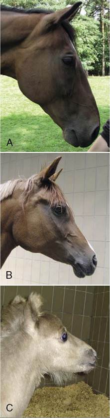
Figure 18-1 Variations in the profile of the equine head. A, The common straight profile. B, The dished Arabian profile. C, The domed contour of the foal.
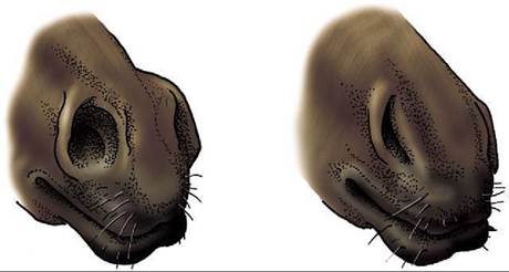
Figure 18-2 Functional variations in the form of the nostril.
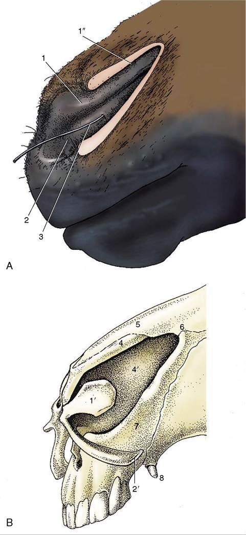
Figure 18-3 A, Left nostril opened laterally to expose nasal diverticulum. B, Nasal cartilages. 1, Alar fold, supported by the lamina (1)' of the alar cartilage; dorsal to the alar fold is the nasal diverticulum (1"); 2, floor of nostril supported by the cornu of the alar cartilage (2')—the floor leads into the nasal cavity; 3, probe in nasolacrimal duct; 4, dorsal lateral nasal cartilage; 4', nasal septum; 5, nasal bone; 6, nasoincisive notch; 7, incisive bone; 8, canine tooth.
manipulation. The dilated nostril is rounded, and the change in form is achieved by apposition of the walls of the diverticulum. The pliancy of the tissues facilitates examination of the nasal vestibule and exposure of the opening of the nasolacrimal duct, which is found on the floor, about 5 cm internal to the entrance and near the mucocutaneous junction. Occasionally the duct has more than one opening.
The entrance to the mouth is small, and the commissure is a little in front of the first cheek teeth (P2). The skin of the lips and adjacent part of the muzzle is sparsely covered by short, fine hairs that impart a velvety texture.
The lips are both mobile and sensitive and are used in the selection and prehension of food. The sensitivity of the upper lip is exploited when a twitch is applied to control a horse during procedures (e.g., injections) elsewhere on the body. The application of acupressure causes the animal to become somewhat sedated while its heart rate is lowered and endorphins are released. It is suggested that the endorphins activate a pain-decreasing mechanism. The lower lip surmounts the chin swelling, which is based on a pad of fatty fibrous tissue.The eyes are prominent and placed to each side of the head, indicating that the horse, like other herbivores, enjoys a panoramic field of vision. Indeed, horses may view almost all around by making only slight movements of the head. This ability to survey widely—perhaps through 330°—is obtained at the expense of the binocular field, which is limited to some 65°. The field of overlap is further reduced by the length and shape of the muzzle, which creates a blind area directly to the front (see Figure 9-1).
The upper and lower eyelids and adjacent skin carry a few scattered tactile hairs. The palpebral skin is thin and, being loosely attached, is thrown into folds when the eye is open. The lid margins carry numerous lashes, longer and more prominent on the upper than on the lower lid (Figure 18-4). The tarsal glands, which open at the junction of the skin with the conjunctiva, number about 50 in the upper lid, rather fewer below, and are clearly visible in palisade formation when the lids are everted. The palpebral conjunctiva is well vascularized, the bulbar part less generously; the bulbar conjunctiva is strongly pigmented toward the corneoscleral region. The third eyelid (Figure 18-5/7) in the medial angle can be exposed in the usual way by pressing on the eyeball through the upper eyelid; a small accessory lacrimal gland is associated with it. The lacrimal caruncle is prominent. The features of the eyeball are considered later (p.
527).A depression caudal to the eye (behind the palpable postorbital bar of bone) is prominent in the animal at rest. It disappears and reappears during feeding in rhythm with the movements of the jaws; the effects are
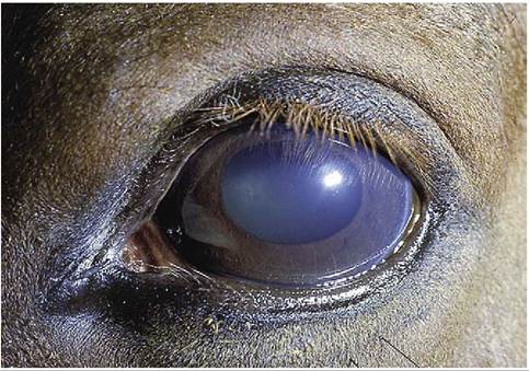
Figure 18-4 Left equine eye; note implantation of eyelashes on lateral side of upper eyelid.
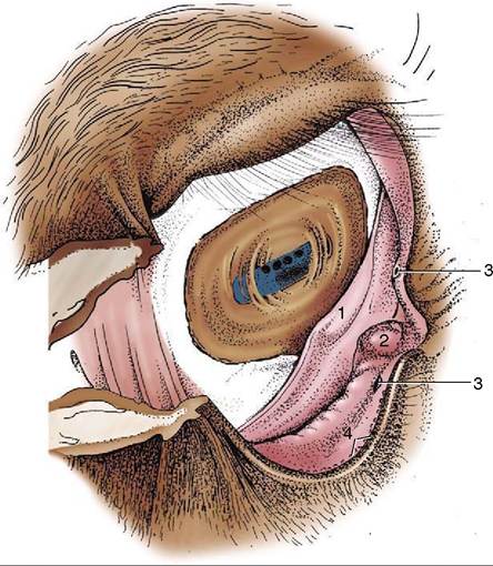
Figure 18-5 The right conjunctival sac. 1, Third eyelid; 2, lacrimal caruncle; 3, lacrimal puncta; 4, openings of the tarsal glands.
due to the displacement of a pad of fat interposed between the temporalis and the periorbita. The fat is depleted in horses in poor condition when exaggeration of the hollow contributes significantly to the haggard appearance.
Deposition of fat above the upper eyelid may produce a conspicuous swelling seen in animals suffering from Cushing disease.
Little need be said concerning the external ears, which are prominent and capable of being swiveled
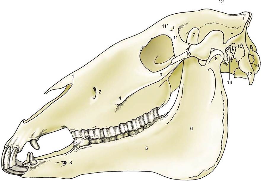
Figure 18-6 Lateral view of the skull. 1, Nasoincisive notch; 2, infraorbital foramen; 3, mental foramen; 4, facial crest; 5, body of mandible; 6, ramus of mandible; 7, coronoid process; 8, condylar process; 9, temporal process of zygomatic bone; 10, zygomatic process of temporal bone; 11, zygomatic process of frontal bone; 11', supraorbital foramen; 12, external sagittal crest; 13, para- condylar process; 14, styloid process; 15, external acoustic meatus; 16, occipital condyle.
when attempts are made to locate the origin of a sound.
Their carriage is also very expressive of emotion.