» The Major Nerves of the Hindlimb
It is only necessary to deal briefly with the course, relations, and distribution of those nerves that extend substantially into the free limb because a general account of the lumbosacral plexus (usually formed by the nerves L4-S2) and its divisions has been presented (pp.
311-313) (Fig. 17.9 and Table 17.2).The femoral nerve (L4-L6) has a very short course within the thigh before it ends by ramifying within the quadriceps femoris, the principal extensor of the stifle and an ancillary flexor of the hip. Shortly before disappearing into this muscle, it detaches the saphenous nerve, which descends subcutaneously over the medial aspect of the limb accompanied by the palpable saphenous artery. Although the saphenous nerve supplies the sartorius, it is largely sensory, serving the skin of the medial surface of the thigh, stifle, leg, and hock (Fig. 17.10). Dysfunction of the femoral nerve paralyzes the quadriceps, resulting in the collapse of the stifle and disabling the entire limb. Compensation is not available. The skin of the medial surface of the limb is deprived of sensation.
The sciatic nerve (L6-S1) crosses the dorsal border of the hip bone to enter the limb together with the caudal gluteal vessels. After passing the dorsocaudal aspect of the hip joint deep to the greater trochanter, where it is susceptible to injury in trauma or surgery of the joint, the nerve and accompanying vessels supply branches to the hamstring muscles. The nerve then continues distally in a central position within the thigh, caudal to the femur and cushioned between the biceps laterally, the adductor, and later the semimembranosus medially (Fig. 17.2/4). At a rather variable point it divides into the common peroneal and tibial nerves that continue the course of the parent trunk until they diverge caudal to the stifle. The sciatic nerve and its peroneal and tibial branches collectively supply the skin of the entire limb distal to the stifle with the exception of the medial strip claimed by the saphenous nerve.
The common peroneal nerve, the more lateral of the terminal divisions of the sciatic nerve, can be palpated in lean dogs where it passes over the lateral head of the gastrocnemius (Fig. 17.5/7). It then dives deeply among the dorsal crural muscles (the extensors of the digits and flexors of the hock), which it supplies. It is continued by superficial and deep (peroneal) branches that enter the paw over the dorsal aspect of the hock; they supply the skin of the dorsal surface. Paralysis of the common peroneal nerve produces slight overextension of the hock and inability to extend the digits, which may be rested on their dorsal surfaces. In time, affected dogs learn to flick their paws forward before putting them down, enabling their limbs to support weight. The dorsal surface of the paw is without sensation.
The tibial nerve passes between the two heads of the gastrocnemius, where it detaches branches to the muscles behind the tibia (the flexors of the digits and extensors of the hock). The depleted nerve, now largely sensory but retaining a small motor component for the intrinsic muscles of the paw, continues distally within the web of skin between the caudal crural muscles and the common calcanean tendon. It crosses the hock beside the deep flexor tendon before branching to supply the plantar structures of the paw. Tibial nerve injuries cause the hock to be flexed and lowered closer to the ground when the limb bears weight. The paralysis of the digital flexors elevates the toes; their plantar aspect is without sensation.
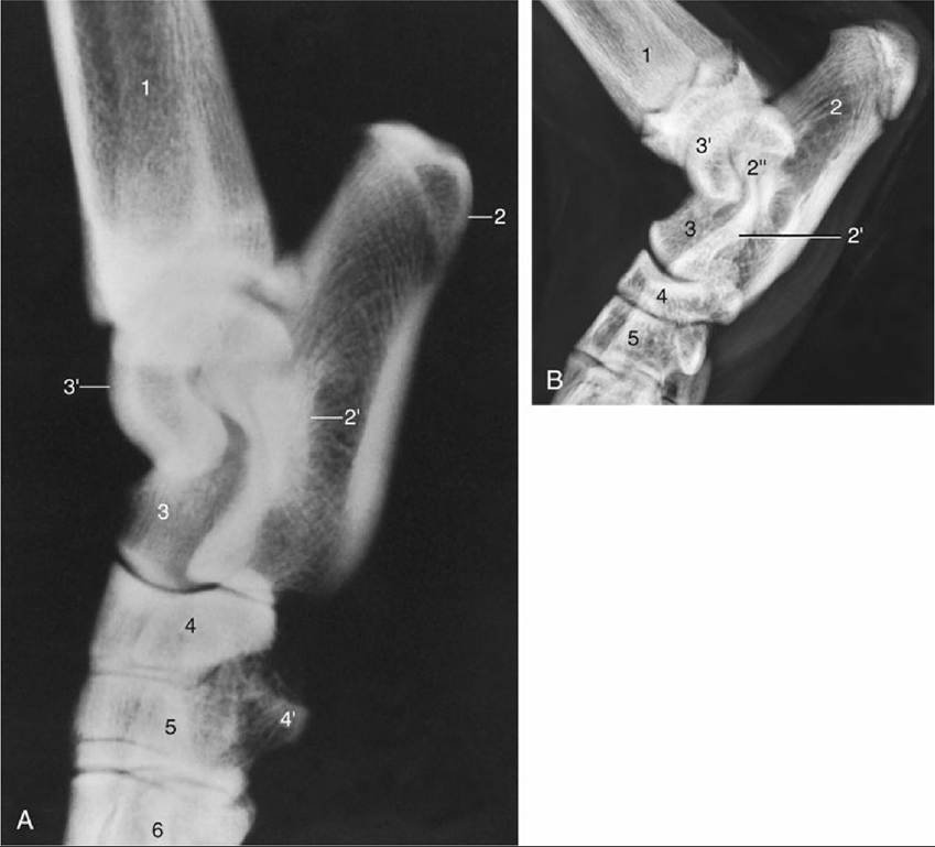
FIG. 17.8 Lateral radiographic views of the (A) canine and (B) young feline hocks. 1, Tibia and fibula; 2, calcaneus; 2', sustentaculum tali; 2", coracoid process; 3, talus; 3', trochlea of talus; 4, superimposed fourth and central tarsal bones; 4', plantar tubercle on fourth tarsal bone; 5, distal row of tarsal bones; 6, metatarsal bones.
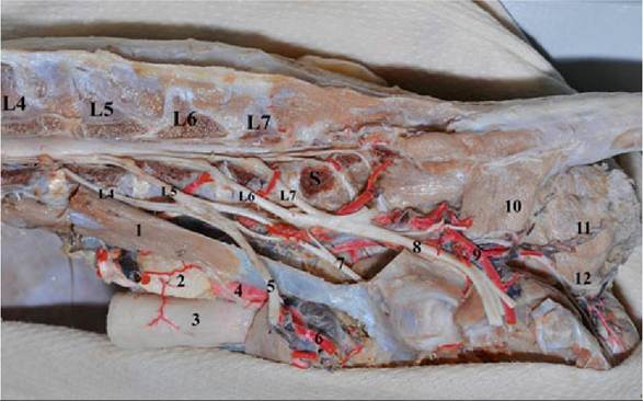
FIG. 17.9 Lumbal plexus. The lumbar vertebral arches and the iliopsoas muscle have been removed. 1. Psoas minor muscle. 2. Medial iliac lymph node. 3. Rectum. 4. External iliac artery. 5. Femoral nerve (Iliopsoas muscle removed). 6. Femoral artery. 7. Obturator nerve. 8. Ischiatic nerve. 9. Caudal gluteal artery and vein. 10. Coccygeus muscle. 11. Caudal rectal artery and vein on the external anal sphincter muscle. 12. Superficial perineal nerve
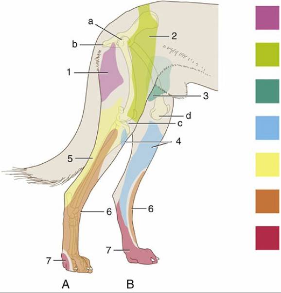
FIG. 17.10 Autonomous zones of the cutaneous innervation on the lateral (A) and medial (B) surfaces of the canine hindlimb. 1, Caudal cutaneous femoral nerve (purple); 2, lateral cutaneous femoral nerve (green); 3, genitofemoral nerve (teal); 4, saphenous nerve (blue); 5, sciatic nerve (yellow); 6, pero-neal nerve (orange); 7, tibial nerve (red). a, Position of greater trochanter; b, ischial tuber; c, lat-eral tibial condyle; d, medial tibial condyle.
Comprehension Check
Describe the anatomy of the hip joint and the anatomic structures that prevent its luxation. Use the cadaver to practice the most efficient surgical approach to the hip joint.
Reviewing the nerve supply to the hindlimb of the dog, describe the impact of dysfunction of each nerve on the stability and locomotion of the limb along with any compensatory mechanisms.
» TABLE 17.2
| Name Origin Insertion Function | |||
| Caudal Muscles of the Thigh and the Hip: all supplied by Sciatic nerve except * by the obturator nerve | |||
| Biceps femoris | All from the ischiatic tuberosity BF has additional origin from sacronιberous ligament | Patella, Tibia | All extend the hip BF and ST extend hock BF extends stifle while ST flexes the stifle |
| Scmitcndiixjsus | Medial side of tibia | ||
| Semimebranosus | Medial side of femur Prox cndoflibia | ||
| Internal obturator | Dorsal pelvic symphysis | Tronchanteric fossa | Rotate the pelvic limb laterally |
| Gcmclli | Ischium caudal to acetabulum | ||
| Quadratus femoris | Caudal ventral ischium | ||
| * External obturator | Ventral pelvic symphysis | ||
| Medial Muscles of thcThigh: All supplied by Obturatorcxccpt * by the femoral nerve | |||
| Gracilis | Pelvic symphysis | Cranial side of tibia | Adduct the limb |
| Pcctineus | Cranial pubic ligament | Medial Surfaccoffcmur | |
| Adductor | Pelvic symphysis | Caudal medial of femur | |
| * Sartorius | Cranial part: crest of Ilium Caudal part: Ilium | Patella Cranial side of tibia | Flcx hip; cranial: extend stifle Caudal: flex stifle |
| Lateral Muscles of the Rump: all SupplicdbyCranial Glutcal nerve except * by the caudal gluteal nerve | |||
| * Superficial Gluteal | Sarcum and Cranial Iliac Spine | 3ri Trochanter | Extend and abduct the hip |
| Middle GIuteaI | Crest ofilium | Greater Tronchanter | |
| Deep Gluteal | Bodyofilium and ischiatic spine | ||
Quadriceps femoris; All heads from proximal fαnur and extend stifle except Rcctus femoris from ilium which flexes hip.
Femoral nerve. Psoas major and Iliacus insert at lesser Ironchanter; Flexes hip; Femoral nerve
CrankjIateral Muscles of the Leg: all supplied by the peroneal nerve
| Cranial tibial | Lateral condyle of tibia | Plantar surface of metatarsal I-Il | All flex tarsus plus LDE extending digits; Cranial tibial rotates paw laterally Peron Iongus rotates paw medially |
| Long digital extensor | Extensor fossa of femur | Distal phalanges of Π-V digits | |
| Peroneus Iongus | Lateral condyle of tibia | 4 a Iaraal + plantar of metatarsals | |
| Caudal Muscles of the Leg: all supplied by the tibia nerve | |||
| Gastrocnemius | Medial & Iat supracondylar tuberosities of femur | Tubercalcanei | Extend tarsus; flex stifle |
| Superficial digital flexor | Lat supracondylar tubil⅛mur | Tubercalcanci and middle phalanges of II-V digits | Extend tarsus; flex digits |
| Deep digital flexor | Tibia and fibula; proximal Caudo-Iateral | Distal phalanges | |
| Popliteus | Lateral condyle of femur | Prox caudal border of tibia | Rotate leg medially |
Arteries: Internal and external ilines; deep femoral; femoral-popliteal-cranial tibial; saphenous; Veins: Medial and lateral saphneous; cranial tibial; femoral