» The Hock and Hindpaw (see also pp. 85, 89-90.)
Inspection of the distal part of the limb reveals the distinctive conformation of the hock; when it is taken apart, there is little external difference between the forepaws and hindpaws beyond the absence of any analogue of the carpal pad.
A dewclaw is commonly present at birth in dogs but is routinely removed at an early age in puppies of many breeds. Duplication of this digit is known to occur in the French Shepherd breeds. Dewclaws of the hindlimb are not found in cats.Although the hock skeleton is complete—there is no suppression or fusion of the standard elements—most bones cannot be individually identified on palpation. The most distinctive feature is the long, rather slender calcaneus, which provides the leverage for the effective extension of the hock. The bone is occasionally fractured by the force exerted by the powerful muscles attaching to its slightly swollen tip. The calcaneus extends a medial process, the sustentaculum tali, over the plantar aspect of the talus, where it may be felt despite being covered by the deep flexor tendon (Fig. 17.7/3'). The more distal tarsal bones lack identifying surface features, but their locations and extents may be deduced after reference to a skeleton or to radiographs. The other prominent surface features of the region are the projections of the tibial and fibular malleoli at the lower limit of the leg and the equally prominent swellings at the proximal ends of the second and fifth metatarsal bones. A long collateral ligament may be traced from the malleolar to the metatarsal thickening on each side of the limb. The extensor tendons can be followed over the dorsal surface of the hock; the retinacula that hold them in place over the distal tibia and again at the proximal end of the metatarsus can also be appreciated in many dogs.
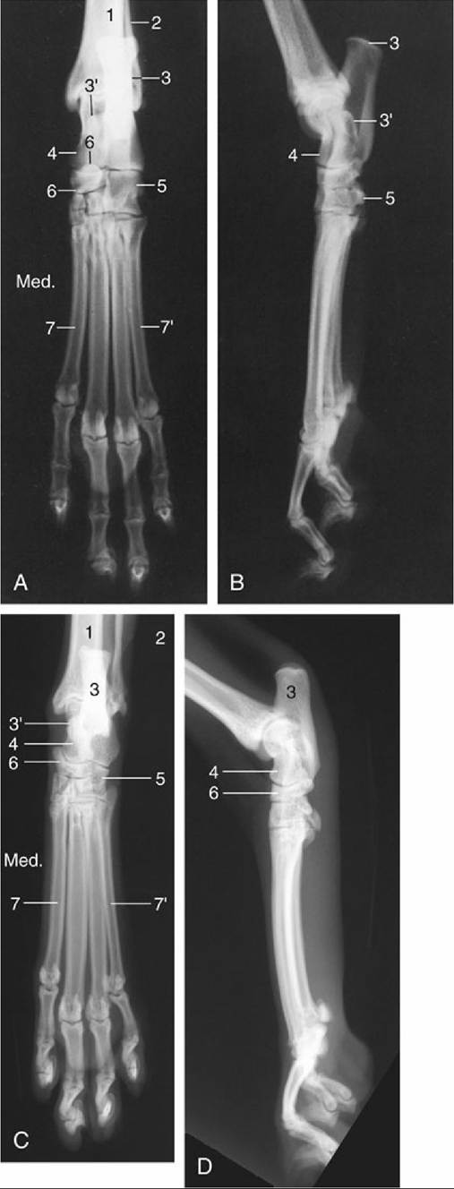
FIG.
17.7 (A) Dorsoplantar (Med., medial) and (B) lateral radiographic views of the canine hocks and hindpaws. (C) Dorsoplantar (Med., medial) and (D) lateral radiographic views of the feline hocks and hindpaws. 1, Tibia; 2, fibula; 3, calcaneus; 3', sustentaculum tali; 4, talus; 5, fourth tarsal bone; 6, centraltarsal bone; 7 and 7', second and fifth metatarsal bones, respectively.
Only the tarsocrural joint is large enough to be punctured in the live animal. This is done on the lateral side just distal to the malleolus; the needle is directed distally toward the lateral surface of the palpable lateral trochlear ridge of the talus.
Similar impressions of the bones and soft structures are obtained on palpation of the hindpaw as on palpation of the forepaw.
Although a complete radiographic examination of the hock calls for exposures in dorsoplantar, mediolateral, and oblique projections, the most useful general picture is obtained from the dorsoplantar view because it permits identification of all the bones, of which some are more easily identified than others because there is considerable superimposition (Fig. 17.7A). Both the talus and the calcaneus are well outlined despite the overlap of the sustentaculum tali. The two bones in the subjacent tier, the fourth and central tarsals (Fig. 17.7/5 and 6), are also generally well outlined, although the mediodistal part of the fourth is superimposed on the third. The second tarsal is clearly shown with the smaller first tarsal superimposed on it. The distal extremities of the tibia and fibula appear closely related in this projection; the gap between them is unexpectedly wide in slightly oblique projections obtained of the cat's hock and is a feature that is occasionally misinterpreted as evidence of luxation.
The lateral projection (Fig. 17.7B) depicts the calcaneus and talus clearly, although they overlap toward the center of the field. The more distal bones are less easily identified in this view, apart from the fourth tarsal bone, which is betrayed by a protuberance on its plantar aspect (Fig. 17.8/4'). Because the central tarsal bone is occasionally dislocated, it is important to note the normal alignment of the dorsal borders of the bones of successive tiers. Two previously unrecorded sesamoid bones have recently been described in the Greyhound at the plantar aspect of the hock about the level of the tarsometatarsal joint. Similar to other sesamoids, these can potentially be misinterpreted as chips fractured from the major bones.
There are no distinctive features of the radiological anatomy of the metatarsal bones and phalanges. The short digital muscles are comparable with those in the front limb.