The Major Vessels
The abdominal aorta and caudal vena cava run the length of the abdomen, partly recessed between the right and left sublumbar muscles.
The abdominal aorta gives rise to paired dorsal lumbar arteries; the last pair originates from the median sacral artery.
Near the second lumbar vertebra in the dog, the phrenicoabdominal trunk emerges branches into the caudal phrenic artery and the cranial abdominal artery, which is also the origin of the adrenal arteries. In the cat the caudal phrenic artery originates from the celiac artery as a single artery. The deep iliac circumflex artery branches off the aorta near the sixth vertebra but may split off the external iliac artery in the dog.The ventral branches of the aorta are the celiac artery, the cranial and caudal mesenteric, the renal arteries, and the ovarian/testicular arteries; sometimes paired adrenal arteries also branch off the aorta. The celiac artery branches off directly after the passage of the aorta through the diaphragm and divides into the hepatic, splenic, and left gastric arteries. The hepatic artery courses to the right of the midline before dividing into three to five branches, which supply the individual liver lobes. After giving off the hepatic branches, the hepatic artery bifurcates into the right gastric and gastroduodenal arteries. The gastroduodenal artery in turn divides into the right gastroepiploic and pancreaticoduodenal arteries.
The cranial mesenteric artery originates one vertebra behind the celiac artery and forms the base of the mesentery. It gives rise to the ileocolic, pancreaticoduodenal, and jejunal arteries in the dog and cat. The renal arteries branch off in the dog ventral to the first and second lumbar vertebrae and in the cat ventral to the third and fourth, and directly caudal to these vessels the genital arteries split off. Ventral to the fifth lumbar vertebrae is the origin of the caudal mesenteric artery, and one to two vertebral bodies more caudal, the external iliac arteries split off to supply the hindlimbs.
The abdominal aorta terminates opposite the seventh lumbar vertebra by bifurcating into right and left internal iliac and middle sacral arteries (Fig. 14.3). The aorta lies in the furrow formed by the left and right iliopsoas muscles.Aortin Thrombus: In both dog and cat, but especially in the cat, the terminal segment of the aorta is commonly the location of a large thrombus, often known as a "saddle” thrombus from its disposition across the division, which may partially or wholly block the three terminal branches. The origin of the thrombus, the degree of obstruction it causes, and the rate at which it developed determine the severity of the clinical signs, which may include complete paralysis of the hindlimbs. Surgical removal of the thrombus has very poor survival rates.
13^
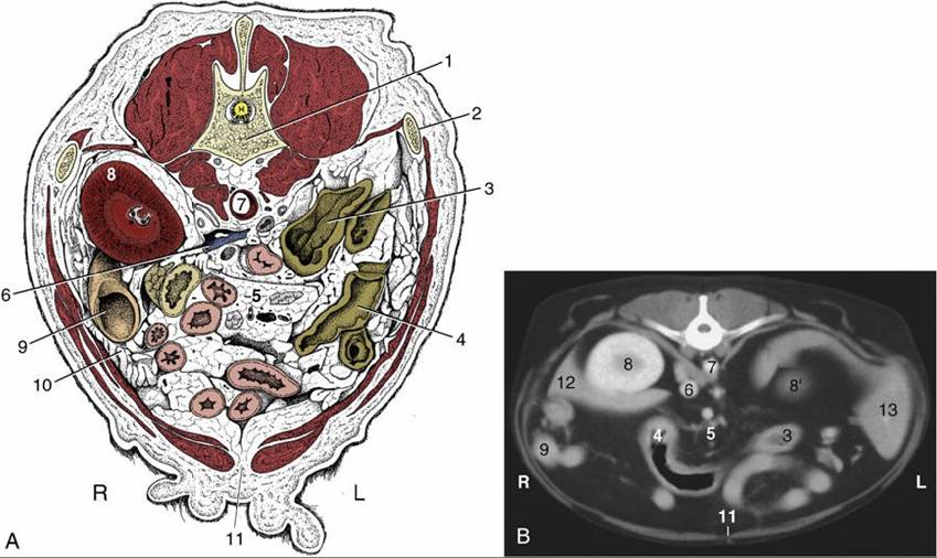
FIG. 14.27 (A) Transverse section of the canine abdomen at the level of the first lumbar vertebra. (B) Corresponding computed tomography (CT) image slightly more caudal than (A); the dog was lying on its back during the CT procedure. 1, First lumbar vertebra; 2, last rib; 3, descending colon; 4, transverse colon; 5, lymph nodes and blood vessels in mesentery; ventral to them is the jejunum; 6, caudal vena cava; 7, aorta, between crura of diaphragm; 8, right kidney; 8', cranial pole of left kidney; 9, descending duodenum and pancreas; 10, greater omentum; 11, linea alba; 12, liver; 13, spleen; L, left; R, right.
The portal vein results from the confluence of the cranial mesenteric, caudal mesenteric, and gastrosplenic veins. In dogs, the portal vein is additionally fed by the gastroduodenal vein, which originates from the merger of the right gastric, right gastroepiploic, and cranial pancreaticoduodenal veins. It has been reported that the contributions to the portal vein in cats are variable and cannot be described on the basis of a common pattern.
Venography of the portal vein (see Fig. 7.45) is occasionally employed to ascertain the existence (and condition) of portosystemic connections. A small intestinal tributary is chosen for the injection. The shunts most commonly revealed connect the portal system with both the caudal caval tributaries at the abdominal roof and the azygos vein within the thorax.