» Lymphatic Structures
The lymph nodes of the abdomen can be divided into a parietal group and a visceral group (Table 14.2). The lumbar aortic lymph nodes, when present, are located along the aorta and vena cava.
They supply the cisterna chyli or the caudal lumbar aortic nodes. The paired hypogastric lymph nodes are small and are located in the angle of the internal iliac and median sacral artery, ventral to the body of the seventh lumbar vertebra. They receive lymph from the thigh, the pelvic viscera, the tail, and a portion of the lumbar region and have efferent vessels to the cisterna chyli. The sacral lymph nodes are positioned ventral to the body of the sacrum but are often not present. They receive afferent vessels from the adjacent musculature and send off efferent vessels to the hypogastric nodes. The deep inguinal or iliofemoral lymph nodes can be found on the ventral surface of the tendon of the psoas minor at its insertion and receive lymph from the pelvic limb. The medial iliac lymph nodes lie between the deep circumflex iliac and the external iliac artery, ventral to the bodies of the fifth and sixth lumbar vertebrae, and they can be 4 cm long in the dog. They receive lymph from all parts of the dorsal half of the abdomen, the pelvis, and the pelvic limb, including that from the genital system and the caudal part of the digestive and urinary system. They also receive lymph from the deep and superficial inguinal, the left colic, sacral, and hypogastric lymph nodes and supply the cisterna chyli. The lymph nodes at the bifurcation of the aorta can be palpated rectally in larger individuals (Fig. 14.32/3).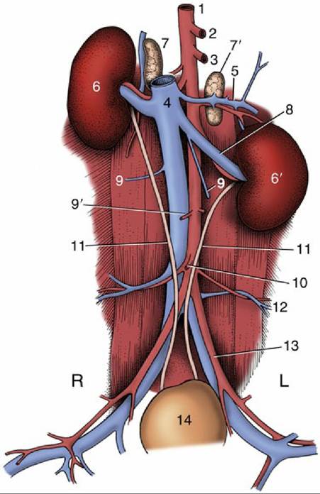
FIG. 14.28 The canine urinary organs and adjacent blood vessels in situ. 1, Aorta; 2, celiac a.; 3, cranial mesenteric a.; 4, caudal vena cava; 5, phrenicoabdominal vessels; 6 and 6', right and left kidneys; 7 and 7', right and left adrenal glands; 8, left renal vessels; 9, ovarian veins.; 9', ovarian arteries; 10, caudal mesenteric artery; 11, ureters; 12, deep circumflex iliac vessels; 13, external iliac vessels; 14, bladder; L, left; R, right.
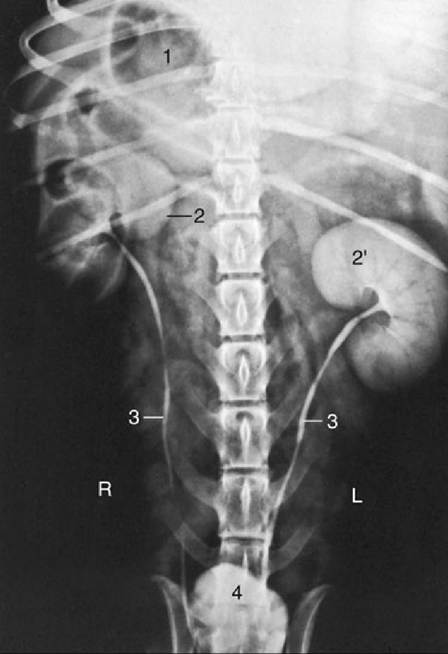
FIG. 14.29 Urogram of a dog. 1, Gas in stomach; 2 and 2', right and left kidneys; 3, ureters; 4, bladder; L, left; R, right.
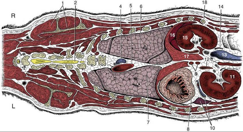
FIG. 14.30 Dorsal section of the canine trunk at the level of the kidneys. 1, Supraspinatus muscle and scapula; 2, spinal cord; 3, sixth and seventh thoracic vertebrae; 4, right azygous vein; 5, thoracic aorta; 6, 7, right and left lungs; 8, fundus of stomach; 9, celiac and cranial mesenteric arteries; 10, splenic vessels and spleen; 11, left kidney; 12, left adrenal gland and abdominal aorta; 13, caudal vena cava; 14, right ureter; 15, right kidney (the right adrenal gland is shown medial to the cranial pole); 16, liver; 17, right crus of diaphragm; 18, last rib; L, left; R, right.
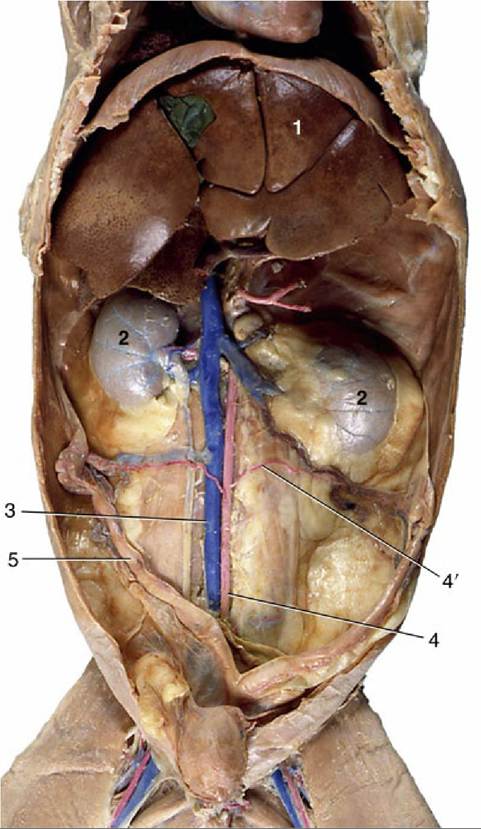
FIG. 14.31 Ventral view of feline abdominal roof. 1, Liver; 2, kidneys (with stellate veins); 3, caudal vena cava (injected); 4, aorta; 4', ovarian artery (injected); 5, uterus.
The visceral lymph nodes are those directly related to the abdominal organs. The gastric lymph node in the lesser omentum near the pylorus is very small and receives lymph from the esophagus, the stomach, the liver, the diaphragm, the mediastinum, and the peritoneum and sends its efferent vessels to the left hepatic or the splenic lymph nodes. The pancreaticoduodenal lymph node is also small, receives lymph from the duodenum, the pancreas, and the omentum, and sends its vessels to the right hepatic or right colic lymph nodes. The hepatic lymph nodes are situated on each side of the portal vein, at 1 to 2 cm from the hilus of the liver. They receive lymph from the stomach, the duodenum, the pancreas, and of course the liver. The three to five splenic lymph nodes along the course of the splenic artery can be 4 cm long in the dog and receive afferent vessels from the esophagus, the stomach, the pancreas, the spleen, the liver, the omentum, and the diaphragm.
The cranial mesenteric lymph nodes are the largest nodes of the abdomen, can be found along the root of the mesojejunum, and receive lymph from the jejunum, the ileum, and the pancreas. The colic lymph nodes in the mesocolon receive afferent vessels from the ileum, cecum, and colon (Table 14.2).The cisterna chyli is an elongated saccular reservoir receiving lymph from the lumbar and mesenteric lymphatic trunks. The cisterna chyli in the dog is located ventral to the first four lumbar vertebrae and dorsal, at the right side, to the aorta and is related to the crura of the diaphragm. The cisterna chyli in cats has a large saccular part dorsal to the aorta and a plexiform part ventral to the aorta and the last thoracic and first three lumbar vertebrae, and it is also closely associated with the diaphragmatic crura.