THE MEDIAL MUSCLES
The medial muscles are disposed in the same three layers as in other species. The superficial layer comprises the gracilis and sartorius (Figure 24-3/8,14). The gracilis exhibits no specific features that require notice.
The sartorius arises from the psoas fascia and the insertion tendon of the psoas minor and gains the thigh by passing through the gap between the caudal margin of the flank and the ilium. It is related to the deep inguinal lymph nodes, where it forms the cranial margin of the femoral triangle. The sartorius inserts on medial structures of the stifle joint, including the condyle of the tibia. Both muscles may adduct the thigh, but the sartorius is probably more important as a hip flexor. The gracilis is supplied by the obturator nerve, the sartorius by the saphenous nerve.The pectineus and adductor constitute the middle layer. The pectineus (Figure 24-3/13) is a small fusiform muscle that arises from the margin of the pubis and inserts on the medial surface of the femur. A part of the tendon of origin is from the contralateral side, and the resulting decussation contributes a transverse strengthening to the prepubic tendon (p. 547). The pectineus is placed to flex the hip and adduct the thigh. It is supplied by the obturator nerve.
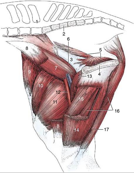
Figure 24-3 Muscles of the thigh, medial view. 1, Last lumbar vertebra; 2, sacrum; 3, shaft of ilium; 4, pelvic symphysis; 5, internal obturator; 6, psoas minor; 7, iliopsoas; 8, sartorius, resected; 9, tensor fasciae latae; 10, rectus femoris; 11, vastus medialis; 12, femoral vessels in femoral triangle; 13, pectineus; 14, gracilis, fenestrated; 15, adductor; 16, semimembranosus; 17, semitendinosus.
The much larger adductor (Figure 24-3/15) fills the space between the pectineus and semimembranosus. It arises from the floor of the pelvis and symphysial tendon and inserts on the caudal surface and medial epicondyle of the femur and the medial collateral ligament of the stifle. Although adduction of the thigh is the primary function, a subsidiary extensor action is possible. Innervation is from the obturator nerve.
The small muscles of the hip—quadratus femoris, gemelli, obturator internus, and obturator externus— are of little importance. The tendon of the obturator internus crosses the margin of the ischium as in the dog. The first three are supplied by the sciatic nerve; while the obturator externus is supplied by the obturator.