THE CRANIAL MUSCLES
This group comprises the quadriceps femoris, which possesses the usual four individually named heads of origin, and the insignificant capsularis.
The four heads of the quadriceps combine in a common insertion on the patella, and the intermediate patellar ligament (Figure 24-4/5) supplies the functional continuation to the tibial tuberosity.
The rectus femoris is a potential flexor of the hip, but the principal action of the group is extension of the stifle. Extension, of course, embraces stabilization of the joint to prevent its collapse when the limb bears weight during the support phase of the stride. It can be observed (and confirmed by palpation) that the muscle appears relaxed when the animal stands quietly. This suggests that, once the patella has been brought into its resting position, no considerable further effort is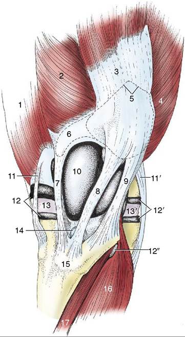
Figure 24-4 The left stifle joint, cranial view. 1, Adductor; 2, vastus medialis; 3, rectus femoris; 4, vastus lateralis; 5, outline of patella; 6, outline of patellar flbrocartilage; 7, 8, 9, medial, intermediate, and lateral patellar ligaments; 10, joint capsule over medial ridge of femoral trochlea; 11, 11', medial and lateral collateral ligaments; 12, 12’, medial and lateral femorotibial joint capsules; 12", recess of 12’ under combined tendon of peroneus tertius and long digital extensor; 13, 13', medial and lateral menisci; 14, distal infrapatellar bursa; 15, tibial tuberosity; 16, long digital extensor; 17, tibialis cranialis.
required of the quadriceps. Quadriceps paralysis is a very severe handicap. The animal is unable to stabilize the stifle; it is also unable to stabilize the hock joint, whose movements are linked to those of the stifle by the reciprocal mechanism (p. 638). The group is supplied by the femoral nerve.