The Medial Muscles
The medial muscles are disposed in the same three layers as in other species. The superficial layer comprises the gracilis and sartorius (Fig. 24.3/8 and 14). The sartorius arises from the psoas fascia and the insertion tendon of the psoas minor and gains the thigh by passing through the gap between the caudal margin of the flank and the ilium.
It is related to the deep inguinal lymph nodes, where it forms the cranial margin of the femoral triangle. The sartorius inserts on medial structures of the stifle joint, including the condyle of the tibia. Both muscles may adduct the thigh, but the sartorius is probably more important as a hip flexor. The gracilis and the sartorius are supplied by the obturator nerve and the saphenous nerve, respectively.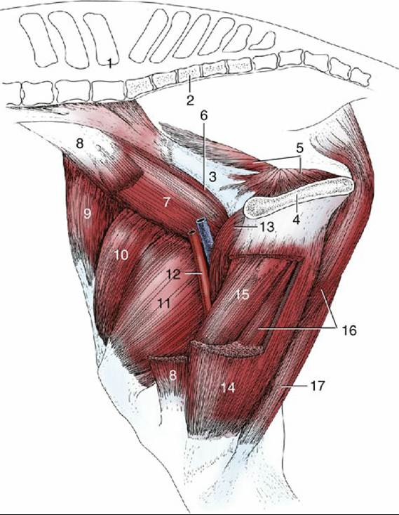
FIG. 24.3 Muscles of the thigh, medial view. 1, Last lumbar vertebra; 2, sacrum; 3, shaft of ilium; 4, pelvic symphysis; 5, internal obturator; 6, psoas minor; 7, iliopsoas; 8, sartorius, resected; 9, tensor fasciae latae; 10, rectus femoris; 11, vastus medialis; 12, femoral vessels in femoral triangle; 13, pectineus; 14, gracilis, fenestrated; 15, adductor; 16, semimembranosus; 17, semitendinosus.
The pectineus and adductor constitute the middle layer. The pectineus (Fig. 24.3/13) is a small fusiform muscle that arises from the margin of the pubis and inserts on the medial surface of the femur. A part of the tendon of origin is from the contralateral side, and the resulting decussation contributes a transverse strengthening to the prepubic tendon (p. 536). The pectineus is placed to flex the hip and adduct the thigh. It is supplied by the obturator nerve.
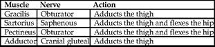
» TABLE 24.3
The Medial Thigh Muscles
The quadratus femoris, gemelli, and obturator internus (supplied by the sciatic nerve) and the obturator externus (supplied by the obturator nerve) are of minor significance.
The much larger adductor (Fig.
24.3/15) fills the space between the pectineus and semimembranosus. It arises from the floor of the pelvis and symphyseal tendon and inserts on the caudal surface and medial epicondyle of the femur and the medial collateral ligament of the stifle. Although adduction of the thigh is the primary function, a subsidiary extensor action is possible. Innervation is from the obturator nerve (Table 24.3).This group comprises the quadriceps femoris, which possesses the usual four individually named heads of origin, and the insignificant capsularis.
The four heads of the quadriceps combine in a common insertion on the patella, and the intermediate patellar ligament (Fig. 24.4/8) supplies the functional continuation to the tibial tuberosity. The rectus femoris is a potential flexor of the hip, but the principal action of the group is extension of the stifle. Extension, of course, embraces stabilization of the joint to prevent its collapse when the limb bears weight during the support phase of the stride. It can be observed (and confirmed by palpation) that the muscle appears relaxed when the animal stands quietly. This suggests that, once the patella has been brought into its resting position, no considerable further effort is required of the quadriceps. Quadriceps paralysis is a very severe handicap. The animal is unable to stabilize the stifle and the hock joint, whose movements are interlinked by the reciprocal mechanism. The group is supplied by the femoral nerve.