» The Stifle Joint
Although generally conforming to the common pattern, the equine stifle also exhibits several important features of distinction. The most remarkable provide the means of "locking" the joint so that one hindlimb may support a disproportionate part of the body weight while standing and allow the other to be rested.
The arrangement is a major component of the passive stay apparatus (p. 625).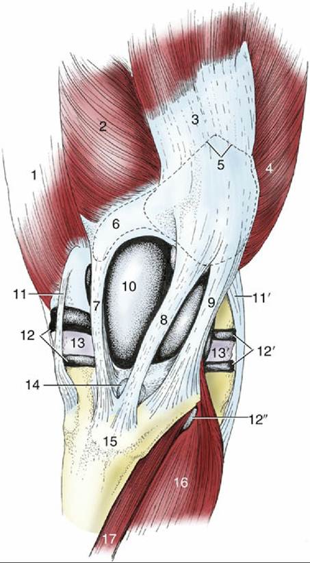
FIG. 24.4 The left stifle joint, cranial view. 1, Adductor; 2, vastus medialis; 3, rectus femoris; 4, vastus lateralis; 5, outline of patella; 6, outline of patellar fibrocartilage; 7, 8, and 9, medial, intermediate, and lateral patellar ligaments, respectively; 10, joint capsule over medial ridge of femoral trochlea; 11 and 11', medial and lateral collateral ligaments, respectively; 12 and 12', medial and lateral femorotibial joint capsules, respectively; 12", recess of 12' under combined tendon of peroneus tertius and long digital extensor; 13 and 13', medial and lateral menisci, respectively; 14, distal infrapatellar bursa; 15, tibial tuberosity; 16, long digital extensor; 17, tibialis cranialis.
The locking mechanism relies on certain peculiarities of the articular surfaces. The femoral trochlea is markedly asymmetrical. The medial ridge is larger than the lateral one and is prolonged proximally to a terminal protuberance that is easily identifiable on palpation (Figs. 24.4/10, 24.5/4, and 24.6/2). The trochlear surface comprises two distinct areas. The larger one, known as the gliding surface, corresponds to the whole trochlea of most species and faces in a predominantly cranial direction; the smaller one, known as the resting surface, forms a narrow shelf above the gliding surface, from which it is sharply angled to face proximally (Fig.
24.7/18). The patella is broadly diamond shaped when viewed from in front (Fig. 24.7B/2). In the fresh state it is extended medially by a parapatellar fibrocartilage (Fig. 24.7/3). The articular surface of the patella is also divided. The more extensive backward-facing area engages with the trochlea during the greater part of the normal range of movement; a narrow strip at the apex is directed distally and makes contact with the femur only at the limit of extension.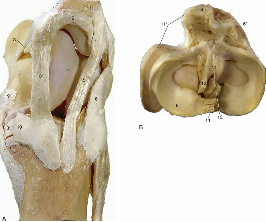
FIG. 24.5 The ligaments of the left stifle joint. (A) Medial view. (B) Proximal view of the left tibia and the menisci. 1, Patella; 2, patellar fibrocartilage; 3, medial femoropatellar ligament; 4, medial ridge of trochlea;
5, intermediate patellar ligament; 6 and 6', lateral and medial patellar ligaments, respectively; 7, lateral collateral ligament, respectively; 8 and 8', lateral and medial menisci, respectively; 9, insertion of semimembranosus; 10, insertion of gracilis and sartorius; 11, meniscofemoral ligament; 11', tendon of popliteus; 12 and 13, cranial and caudal cruciate ligaments, respectively; 14, intercondylar eminence.
The horse has three patellar ligaments joined by a retinaculum in which the insertion tendons of several thigh muscles merge. The intermediate ligament (Fig. 24.4/8), the homologue of the single structure of the smaller species, runs from the apex of the patella to the tibial tuberosity. The lateral and medial ligaments run from the angles of the patella or, more accurately where the medial one is concerned, from the parapatellar cartilage. The three ligaments are thus quite widely separated at their origins but converge distally and insert close together. The gap between the proximal parts of the medial and intermediate ligaments is especially wide and is occupied by the medial ridge of the trochlea (Fig. 24.4/10).
The patella slides up and down over the femoral trochlea during the greater part of the normal movement of the joint.
Only in extreme extension, as momentarily during the support phase of a walking stride, do the resting surfaces engage. The resting position is also adopted when the animal is standing squarely with its weight evenly distributed over the two hindlimbs. This is easily verified on palpation, and it can be found that the medial ligament then runs even with the edge of the corresponding ridge of the trochlea. This position is maintained without the assistance of the main extensor (quadriceps femoris) of the stifle but does require some effort on the part of the muscles that converge on the medial and lateral patellar ligaments: the biceps and tensor fasciae latae laterally and the gracilis and sartorius medially. The position is unstable and the patella is easily dislodged; it then slips back onto the gliding surface of the trochlea.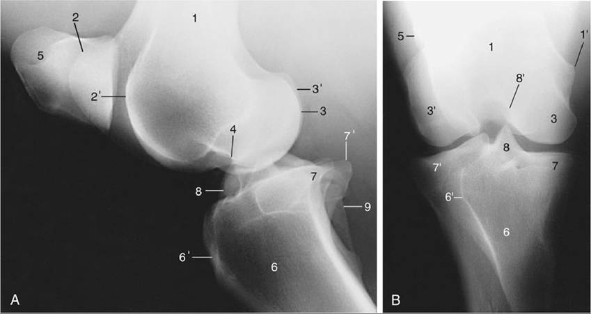
FIG. 24.6 (A) Lateral and (B) caudocranial radiographs of the stifle joint. 1, Femur; 1', medial epicondyle; 2 and 2', medial and lateral ridges of the trochlea, respectively; 3 and 3', medial and lateral condyles, respectively; 4, extensor fossa; 5, patella; 6, tibia; 6', tibial tuberosity; 7 and 7', medial and lateral condyles, respectively; 8, intercondylar eminence; 8', intercondylar fossa; 9, fibula.
The stifle joint cavity is capacious, and its division into compartments is relatively complete. The extensive femoropatellar compartment is mainly contained between the femur, the patella, and the quadriceps. The part distal to the patella is more accessible, though separated from the patellar ligaments (and retinaculum) by a thick cushion of fat. It communicates with the medial femorotibial compartment in the large majority of horses but with the corresponding lateral compartment in far fewer (perhaps 25%). The partition between the medial and lateral compartments is almost always imperforate. The inconstancy of these arrangements should lead to an assumption that any infection spreads readily among the three compartments, while therapeutic substances should be separately injected into each.
The injections into the stifle joint require familiarity with the disposition of the ligaments and the ability to recognize them on palpation. The medial collateral ligament can be picked out close to its origin from the femoral epicondyle and provides a convenient landmark in puncture of the medial femorotibial compartment. The needle is introduced close to its cranial border, between it and the medial patellar ligament (Figs. 24.4/7 and 11 and 24.5/6' and 7'). The lateral collateral ligament is palpable along its whole length but is most easily found close to its insertion on the head of the fibula. The lateral femorotibial compartment is punctured between the lateral patellar ligament and the more cranial, and also palpable, tendon of origin of the long digital extensor (Fig. 24.4/11' and 16). The femoropatellar compartment is also easily entered from the cranial side between the middle and the medial or the middle and the lateral patellar ligaments (Fig. 24.4/9). Alternatively, this compartment can be approached from the lateral side by inserting a needle behind the lateral patellar ligament.
Stifle Joint: Osteochondrosis, a developmental cartilaginous disease, is the most common disease of the stifle joint. Osteoarthritis may develop secondary to some other pathology of the stifle or may be a primary manifestation. Meniscus tears are the most common soft tissue injury in the stifle.