THE MEDIASTINUM
The heart divides the mediastinum into the familiar parts (Figure 20-8/4,4').
The cranial part is markedly asymmetrical; it attaches to the left first rib and gradually shifts to reach a more or less median situation directly in front of the heart.
The dorsal part is thick, the ventral part much thinner, especially after the thymus has regressed. The dorsal part occupies about half the transverse diameter of the thorax and includes the esophagus and trachea, the brachiocephalic trunk and cranial vena cava with their respective branches and tributaries, the cranial mediastinal lymph nodes, the thoracic duct, and the phrenic, vagus, and sympathetic nerves. The interstices between these structures are occupied by fat, sometimes present in large amounts. The thymus is the sole content of the ventral portion.The ventral part of the middle mediastinum is very broad because it contains the heart and pericardium
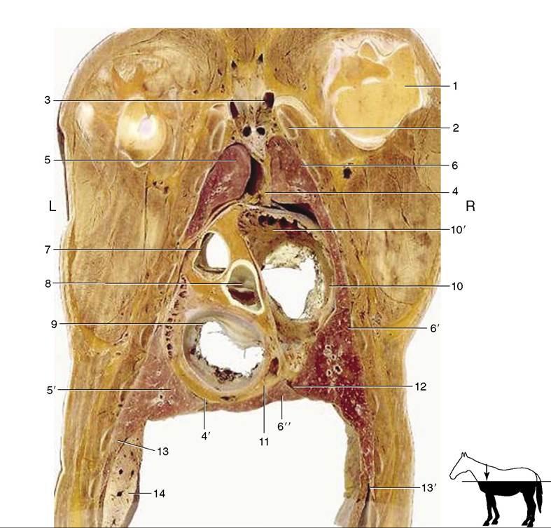
Figure 20-8 Dorsal section of the thorax at the level of the atrioventricular valves. 1, Head of humerus; 2, first rib; 3, formation of cranial vena cava; 4, 4’, cranial and caudal mediastinum; 5, 5’, cranial and caudal lobes of the left lung; 6, 6', 6", cranial, caudal, and accessory lobes of the right lung; 7, pulmonary valve; 8, aortic valve; 9, left atrioventricular valve; 10, right atrioventricular valve; 10', right auricle; 11, coronary sinus; 12, plica venae cavae; 13, diaphragm; 13', costodiaphragmatic recess; 14, part of the liver.
(Figure 20-7). The dorsal part is paper-thin except where it contains the esophagus, the continuation of the trachea to its bifurcation, the aorta, and certain nerves (including vagal branches).
In lateral view the caudal mediastinum is triangular (Figure 20-3/4). It is divided into two parts by adhesion between the lungs about and caudal to their roots. The ventral part, whose sole occupant is the left phrenic nerve, is diverted far to the left before it merges with the pleura covering the diaphragm (Figure 20-4/6). The dorsal part is thin except where it encloses the esophagus and aorta.
Except in foals, small openings in the mediastinum place the two pleural cavities in communication. The mediastinum is very fragile, and exposure during dissection inevitably increases the number of visible openings, which leaves it unclear whether any were present when the thorax was intact and suggests that the mediastinum might be an ineffectual partition. However, small openings in the thoracic wall such as are made for the purpose of thoracoscopy (when the influx of air can be controlled) result in incomplete unilateral pneumothorax and are survived without obvious adverse effects.