The Motility Functions of the Cecum and Colon Retain Material for Fermentation and Separate Particles by Size
The functions of the equine hindgut in maintaining fermentation are similar to those of the rumen: favorable conditions must be maintained to support optimal fermentation. As in the rumen, these conditions are (1) substrate supply, (2) control of pH and osmolality, (3) anaerobiosis, (4) retention of fermenting material, and (5) continual removal of waste products and the residue of spent fermentation substrate.
Separation of fermenting material from residue appears to be accomplished by selective retention of particles according to size, just as in the rumen; however, the means by which the cecum and colon accomplish size separation and discriminate passage are quite different from those of the forestomach. Anatomical characteristics and motility patterns in the cecum and colon are responsible for selective retention of long particles, allowing sufficient exposure for microbial digestion to occur. In general, the fermentative digestive process in the horse is not as efficient as that in the ruminant, and digestible energy values for forages are usually lower for horses than for cattle.Before the motility of the equine cecum and colon is discussed, a brief review of the anatomy of the equine hindgut is important. Figure 31-13 shows the equine digestive system, separated from its mesenteric attachments and laid out in linear fashion. The hindgut commences with the cecum, which is separated from the large colon by a well-defined orifice. The large colon is folded on itself three times, forming four major anatomical divisions: the right ventral and left ventral and the left dorsal and right dorsal colon segments. Ingesta enter the right ventral colon and course to the left ventral colon, from which the material enters the left dorsal portion through the pelvic flexure. From the left dorsal colon, material moves to the right dorsal colon before entering the small colon.
(Anatomy textbooks describe the arrangement of the large colon in the abdomen.) For the purposes Ofphysiological study, the reader should note in Figure 31-13 the tremendous size and volume of the cecum and colon compared with the small intestine.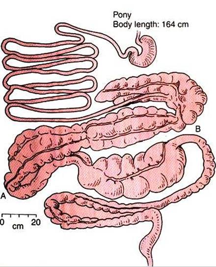
FIGURE 31-13 ■ Equine gut. Note the tremendous development of the colon compared with the small intestine. Note also the relative areas of constriction at the junctions of the ventral and dorsal colons (A) and the large and small colons (B). (From Stevens CE: Comparative physiology of the digestive system. In Swenson MJ, editor: Dukes' physiology of domestic animals, ed 9, Ithaca, NY, 1977, Cornell University Press.)
The differences in diameter that occur throughout the colon should also be noted, particularly the reductions in diameter that occur at the pelvic flexure and at the junction of the large and small colons. The saclike evaginations that occur in the wall of the cecum and most segments of the colon are called hαustrα. Functionally, the equine hindgut can be divided into four sections: cecum, ventral colon, dorsal colon, and small colon.
Ingesta reach the cecum after a relatively short time in the stomach and small intestine. A large portion of soluble ingesta usually reaches the cecum by 2 hours after ingestion, whereas solids take somewhat longer, depending on particle size and consistency. The material in the cecum and throughout the large colon has a high water content and a slurrylike consistency.
The majority of cecal motility is of a mixing nature, with frequent low-amplitude contractions that transport ingesta from haustrum to haustrum and back in a mixing pattern. The mixing action of the cecum maintains the cecal contents in a homogeneous state. About once every 3 to 4 minutes, there is a strong contraction of cecal muscles in a massmovement type of action (see Chapter 28 for a description of mass movement) in which the body and apex of the organ shorten and constrict, lifting ingesta into the base.
Constriction of the base forces material through the cecocolic orifice and into the right ventral colon. The motility pattern functionally separates the cecum from the ventral colon, with no apparent mixing of contents between the two hindgut segments. Thus, there is no retrograde flow of material from the colon to the cecum, so the composition of ingesta in these two organs usually differs somewhat.Three types of motility patterns exist in the right and left ventral colon: Iiaustral segmentation, propulsive peristalsis, and retropulsive peristalsis. Segmentation serves a mixing function that aids in promoting fermentation and bringing VFAs in contact with the mucosa for absorption. Mixing occurs throughout the ventral colon, and the right and left segments may be regarded as one functional unit with homogeneous ingesta. Propulsive activity, or aboral peristalsis, in the ventral colon originates near the cecum and appears to occur as a continuation of the cecal mass movements. Peristaltic activity in the proximal ventral colon propels ingesta distally into the left ventral colon. In the left ventral colon, retropulsive or antiperistaltic movements resist the flow of ingesta and result in the retention of material in the ventral colon, allowing time for microbial digestion and preventing the washout of microbes. In addition, the retropulsive actions of the left ventral colon aid in creating differential flow rates of liquid and particulate matter through the colon. The antiperistaltic motility appears to originate from a pacemaker in the pelvic flexure, the area of restricted diameter where the left ventral and left dorsal colons meet.
1 he motility of the ventral colon can be roughly compared with that of the stomach, with the pelvic flexure and distal left ventral colon acting as the pylorus and antrum, respectively. The pumping action of cecal mass movements, combined with the propulsive action of the proximal ventral colon, continually moves ingesta toward the pelvic flexure.
In the distal ventral colon, however, antiperistaltic activity and the narrow diameter of the pelvic flexure impede the movement of material, causing it to be retained in the ventral colon. The squeezing action of the pelvic flexure mimics the action of the pylorus in selectively retaining relatively large particulate matter while allowing liquid and small particles to pass. As particle size is reduced by fermentative action and the mixing activity of the colon, particles eventually become small enough to flow with the fluid phase and leave the colon. The action of the pelvic flexure is not as efficient as that of the pylorus, and some large particles do escape the ventral colon. In addition, there are periods during which propulsive movements occur in the left ventral colon and pelvic flexure. These factors allow the movement of particulate matter into the left dorsal colon.The actions of the dorsal colon appear to mimic those of the ventral colon. Impedance to ingesta flow is created by the size restriction at the junction of the right dorsal colon and small colon. In addition, retropulsive motility may originate in the area of the distal right dorsal colon, near the junction with the small colon. These actions tend to impede the movement of ingesta through the dorsal colon, subjecting the material to another round of fermentative digestion, as occurred in the ventral colon. The delay in the flow of ingesta created by the combined actions of the ventral and dorsal colons results in significant retention of material, with most particulate matter taking from 24 to 96 hours to pass the large colon. The efficiency of the large colon in retaining and separating ingesta of different particle sizes can be understood from Figure 31-14.
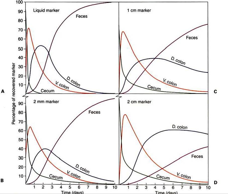
FIGURE 31-14 Retention of liquid and particles of various sizes in the compartments of the equine large intestine. Marker liquid (A) and marked particles of various sizes (B, 2 mm; C, 1 cm; D, 2 cm) were placed in the cecum of ponies, and the distribution of marked materials among colon segments was measured at 2-hour intervals.The lines of the graph were mathematically fitted to the data.
Each line indicates the percentage of marker in a given segment at any time. Note in graph A that at 7 days after infusion, almost all the liquid marker had been recovered in the feces, with little or none remaining in the intestinal segments. Increasing particle size has a relatively small effect on the movement of particles out of the cecum. In contrast, as particle size increases, there is significant retention of material in the colon and slow passage to the feces. D, Dorsal; V, ventral. (From Argenzio RA, Lowe JE, Pickard DW, et al: Digesta passage and water exchange in the equine large intestine. Am J Physiol 226:1035, 1974.)Understanding the motility of the equine colon is important because problems of colon impaction in horses are common. Impactions usually occur near or within the pelvic flexure, probably because the pelvic flexure is a site of flow restriction and differential flow of solid and liquid material. One can easily appreciate how the normal motility pattern could allow solid material to accumulate in this area and cause obstructions.
Although general understanding of small colon motility is limited, it appears to consist primarily of segmentation and propulsion. The characteristic fecal balls of horses are formed by segmentation within the small colon.