THE MOUTH
The term mouth (os, gen. oris) designates not only the cavity and its walls but also the accessory structures that project (teeth, tongue) and drain (salivary glands) into it. The mouth has as its main functions the prehension, mastication, and insalivation of food.
It may also play a role in aggression and defense, while in ourselves it is important in the formulation of the sounds of speech. In most species it functions as an airway when flow through the nose is impaired.The mouth (oral) cavity is entered between the lips and continues into the pharynx (Figure 3-3) through a caudal narrowing at the level of the palatoglossal arches (see further on). It is divided by the teeth and margins of the jaws into an outer vestibule, bounded by the lips and cheeks externally, and the central mouth cavity proper. When the mouth is closed, these divisions communicate through gaps behind and between the teeth. The vestibule extends caudally toward the ramus of the mandible and the masseter muscle. The proportion of its walls formed by the lips varies with feeding habits; a wide gape is necessary in species that feed greedily or use their teeth to seize prey or in fight, whereas a smaller opening suffices in most herbivores and rodents.
Diet and feeding habits also determine the form of the lips (labia oris). In some species, such as the horse, the lips are employed in collecting food and introducing it to the mouth; for this purpose they must be both sensitive and mobile. When other parts are more important in prehension the lips can be less mobile and reduced in size (e.g., cat) or thickened and insensitive (e.g., ox). The lips of the dog are extensive but thin, and although they can be drawn back from the teeth, they are not capable of other purposeful movements. Lip posture is an important factor in communication in this species and can signal aggressive intent or submission.
In newborn animals the lips form the seal about the teat that is necessary for successful sucking.The lips are composed of skin, an intermediate layer of muscle, tendon, and glands, and the oral mucosa. The skin and mucosa usually meet along the margin of the lips, though the boundary can be displaced in either direction. The muscles that make up the greater part of the lips belong to the mimetic musculature, which is the field of the facial nerve. They include an orbicular muscle encircling the opening and, with some species variation, others that raise, depress, and retract the lips. Small salivary glands are scattered among the muscle bundles below the mucosa, especially toward the angles (commissures) where the two lips meet.
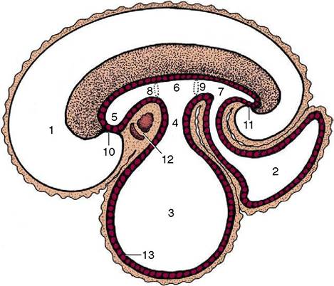
Figure 3-2 Sagittal section of an early embryo. Part of the yolk sac is taken into the body in the folding process. 1, Amniotic cavity; 2, allantoic cavity; 3, yolk sac; 4, stalk of yolk sac; 5, foregut; 6, midgut; 7, hindgut; 8, cranial intestinal portal; 9, caudal intestinal portal; 10, oral plate; 11, cloacal plate; 12, heart and pericardial cavity; 13, endoderm.
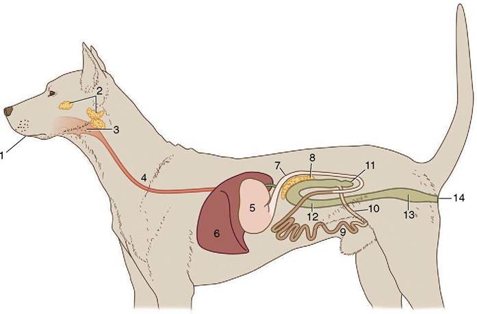
Figure 3-1 Schematic representation of the digestive apparatus in the dog. 1, Mouth; 2, salivary glands; 3, pharynx; 4, esophagus; 5, stomach; 6, liver; 7, duodenum; 8, pancreas; 9, jejunum; 10, ileum; 11, cecum; 12, colon; 13, rectum; 14, anus.
There is rarely anything remarkable in the arrangement of the lower lip. In the dog it is rather loose but fastened to the lower jaw at the level of the canine tooth and has a thin, serrated margin. Modifications of the upper lip are more frequent. Sometimes a median naked area is present continuous with the modified skin around the nostrils. The extensive moist and glandular nasolabial plate of the ox and the rostral disk of the pig are good examples of this.
The area of modified skin is often much narrower and may be divided by a median groove (philtrum) as in the dog. Dog breeders refer to this modified region as the “nose leather” (see Figure 3-3). In man and in the horse a hairy integument extends across the entire upper lip.The cheeks (buccae), which tend to be most capacious in herbivores, have a similar structure. The principal support is the buccinator muscle, which has the important function of returning to the central cavity any food that has escaped into the vestibule. There are additional salivary glands, sometimes aggregated in quite large masses: the zygomatic gland of the dog (see Figure 3-12∕the tongue forward. The genioglossus arises more dorsally than the geniohyoideus and first runs back below the floor of the
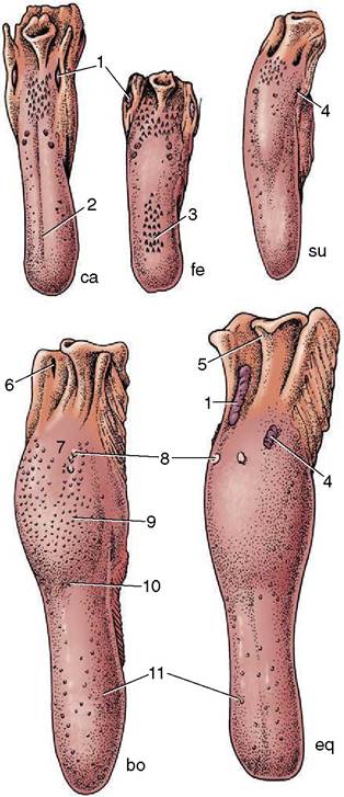
Figure 3-9 Dorsal view of the tongue and epiglottis of the dog (ca), cat (fe), pig (su), cattle (bo), and horse (eq). 1, Palatine tonsil; 2, median groove; 3, filiform papillae; 4, foliate papillae; 5, epiglottis; 6, tonsillar sinus; 7, root of tongue; 8, vallate papillae; 9, torus linguae; 10, fossa linguae; 11, fungiform papillae.
mouth before dividing into bundles that fan upward in the sagittal plane. Those bundles that turn forward to the apex of the tongue retract this part; those that pass toward the root draw the whole tongue forward. The middle group passes toward the upper surface (dorsum), which it may depress. The other two muscles arise from the hyoid apparatus. The hyoglossus takes origin from the basihyoid and runs forward, lateral to the genioglossus; the styloglossus takes origin from the stylohyoid but farther to the side. Both draw the tongue back but in rather different fashions; the styloglossus also tends to elevate it. The intrinsic muscle is disposed in bundles that run longitudinally, transversely, and vertically (see Figure 4-2). Simultaneous contraction of the transverse and vertical bundles stiffens the tongue.
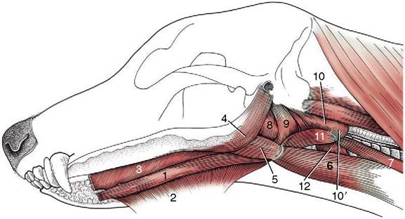
Figure 3-10 Muscles of the tongue and pharynx of the dog. 1, Geniohyoideus; 2, mylohyoideus; 3, genioglossus; 4, styloglossus; 5, hyoglossus; 6, sternohyoideus; 7, sternothyroideus; 8,9, hyopharyngeus (two parts); 10, thyropharyngeus; 10', cricopharyngeus; 11, thyrohyoideus; 12, cricothyroideus.
The muscle bundles are interspersed with considerable amounts of fat, which is an arrangement that imparts a unique consistency and flavor to the cooked tongue. This fat is very resistant to mobilization in starvation.
In the dog, alone among the domestic species, the ventral part of the tongue contains a prominent fibrous condensation, the lyssa, easily recognized on palpation. A fibrous septum that extends from this is responsible for the conspicuous median groove on the upper surface.
The innervation accurately reflects the origin of the tongue as an unpaired swelling of the pharyngeal floor (see Figure 3-58, C) that is later extended by contributions from the ventral parts of the adjacent pharyngeal (branchial) arches. The mucosa retains a sensory innervation from the corresponding arch nerves. The lingual branch of the mandibular nerve is responsible for general sensation over the rostral two thirds of the tongue; the chorda tympani, a branch of the facial nerve, is responsible for the special sensation of taste in the same area. Both general and special sensation of the root region are the responsibility of the glossopharyngeal and, to a small extent, the vagus nerves. The extrinsic and intrinsic muscles are all supplied by the hypoglossal nerve, although it is probable that the sensory fibers emanating from spindles and other receptors in these muscles travel mainly in the lingual nerve.
Relatively little of the floor of the mouth is left accessible rostral and lateral to the attachments of the tongue. The largest free area lies ventral to the apex, behind the incisor teeth. The mucosa here covers the incisive part of the mandible directly, but elsewhere it lies on muscle and the floor is yielding. The most prominent features are fleshy protuberances or caruncles behind the central incisors; these carry the common openings of the mandibular and major sublingual salivary ducts (see Figure 3-3). In some species, much smaller serial elevations to each side of the frenulum mark the openings of the lesser ducts of the sublingual gland. The mylohyoideus muscle passes below the mucosa and tongue from a linear attachment on the medial aspect of the mandible to meet its fellow of the other side in a median raphe; the two together suspend the tongue in a muscular hammock (see Figure 3-21/4). This muscle is supplied by the mandibular nerve and plays an important part in initiating swallowing (p. 121).