THE SALIVARY GLANDS
Numerous salivary glands drain into the oral cavity. Their secretion, the saliva, keeps the interior of the mouth moist, and when mixed with food, saliva facilitates mastication.
When the food is eventually formed into a bolus for swallowing, the saliva lubricates its passage.Small salivary glands have been mentioned as features of the lips, cheeks, and tongue; others are present in the soft palate, pharynx, and esophagus. Although individually unimportant, their collective contribution must be considerable. However, most saliva comes from certain larger glands situated at a greater distance from the mouth cavity into which they drain through longer ducts (Figure 3-11). Unlike the minor glands, which mostly produce a mucous secretion, some of these major glands produce a more watery (serous) fluid containing the enzyme ptyalin, which plays a minor role in carbohydrate digestion.
The parotid gland, which is purely serous in most species (though not in the dog), obtains its name from its relationship to the ear, being molded around the ventral part of the auricular cartilage (Figure 3-12). In the dog it is small and confined to the vicinity of the
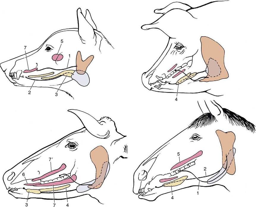
Figure 3-11 The major salivary glands of the dog, pig, cattle, and horse. Orange: parotid gland; white: mandibular gland; yellow: sublingual glands; red: buccal glands. 1, Parotid duct; 2, mandibular duct; 3, compact (monostomatic) part of sublingual gland; 4, diffuse (polystomatic) part of sublingual gland; 5, dorsal buccal glands (zygomatic gland in the dog); 6, middle buccal glands; 7, ventral buccal glands; 7, middle buccal gland.
cartilage. Because the serous parotid secretion is important in moistening and softening food, the gland is larger and the flow more copious in herbivores.
In these species the parotid gland extends rostrally onto the masseter muscle, ventrally toward the angle of the jaw, and caudally toward the atlantal fossa. In all species it is enclosed within a fascial covering that sends trabeculae inward to divide the gland into obvious lobules.The major collecting ducts run within these trabeculae and eventually join to form a single duct that leaves the cranial aspect. In the dog this duct takes the shortcut across the lateral surface of the masseter to open into the vestibule of the mouth opposite the fourth upper premolar tooth. In the large domestic animals the duct takes the longer but more protected route medial to the angle of the jaw and winds below the mandible to enter the face along the rostral margin of the masseter.
The mandibular gland produces a mixed mucous and serous secretion. Generally smaller than the parotid, it is more compact and is placed close to the angle of the jaw. It is a moderately large, very regular ovoid structure in the dog. It too is much larger in herbivores, in which it has a deeper position. This gland also drains by a single large duct that runs ventral to the mucous membrane of the floor of the mouth, close to the frenulum of the tongue, to open on the sublingual caruncle.
The sublingual gland is also commonly mixed and sometimes consists of parts: one is compact (monostomatic) and drains by a single duct, and the other is diffuse (polystomatic) and opens by several small ducts. In the dog the compact part fits over the rostral extremity of the mandibular gland, which it appears to continue. The duct that leaves this part runs close to the mandibular duct and discharges alongside this or through a common opening. The diffuse part,
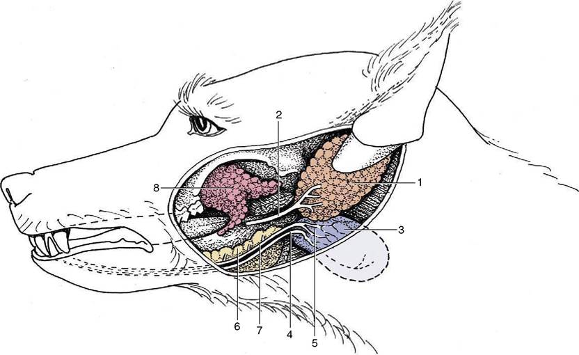
Figure 3-12 The salivary glands of the dog. 1, Parotid gland; 2, parotid duct; 3, mandibular gland; 4, mandibular duct; 5, caudal part of compact sublingual gland; 6, rostral part of compact sublingual gland; 7, major sublingual duct; 8, zygomatic gland.
the only part present in the horse, is a thin strip lying below the mucosa of the oral floor; its many ducts open beside the frenulum.
The flow of saliva is normally continuous, although the rate is influenced by many factors. It is depressed by anxiety or fear and may be wholly suspended when the body is dehydrated: the resulting dryness of the mouth contributes to the sensation of thirst. It is increased when substances—even inedible ones—are introduced into the mouth, although food is most effective, as was demonstrated by the classic experiments of Pavlov. Events indicating that feeding is imminent are also effective. The rate of secretion is controlled by the innervation. The salivary glands receive both sympathetic and parasympathetic supplies, the latter being vastly more important. The parasympathetic fibers come from the two salivatory nuclei of the brainstem and first travel in the facial and glossopharyngeal nerves; later the fibers pass into various branches of the trigeminal nerve that convey them to their destinations. The preganglionic fibers synapse close to the gland, and the postganglionic fibers terminate in direct contact with the secretory cells. Stimulation is followed by copious flow accompanied by vasodilation. Sympathetic stimulation produces vasoconstriction, which slows the rate of production and alters the composition of the saliva.
In addition to its cleansing, lubricant, and digestive functions, saliva serves as a route for the excretion of certain substances, some of which may accumulate as a deposit (tartar) on the teeth.