THE MOUTH
The stomodeum, carried ventrally in the folding process, comes to lie between the swelling of the forebrain dor- sally and that over the developing heart ventrally. The oral membrane soon breaks down; with its disappearance it is no longer possible to recognize the extent of the ectodermal contribution to the lining of the mouth.
The mouth is built up by the forward growth of certain processes that appear around the margins of the oral plate. Dorsally, a frontal process appears as the result of a spurt in growth of the paraxial mesoderm around the forebrain. Laterally and ventrally, the margin is formed by the mandibular arch, the first of the thickenings (see further on) that develop in the mesoderm lateral to the presumptive pharynx.
The frontal process is initially a simple prominence. Soon bilateral thickenings, olfactory placodes, appear in the covering ectoderm immediately bounding the oral depression. These placodes sink below the surface when growth of the surrounding mesoderm throws up a rim around each. The rim has the form of a horseshoe with a ventral interruption leading to a groove extending to the mouth. The interruption divides the lateral and medial parts of the rim, which are known hereafter as the lateral and medial nasal processes. The mandibular arches also expand and grow toward each other at this time; they soon fuse ventral to the oral depression, forming the continuous shelf of the lower jaw and mouth floor. In addition, the upper end of each mandibular arch detaches a maxillary process that extends forward between the frontal and mandibular processes to enclose the mouth laterally. The various swellings gradually merge.
The depressions in which the olfactory placodes are contained originally communicate with the oral cavity, but these connections are lost when the placodes sink more deeply within blind pits, the nasal fossae, that now excavate the upper jaw.
The tissue that remains between these pits and the mouth constitutes the primary palate. Communication between nose and mouth is regained when the pits eventually break through into the mouth cavity at two openings known as the primitive choanae (Figure 3-57). The disruption is considerable, and only the most rostral part of the primary palate survives.The definitive nasal cavities arise from a fresh subdivision of the temporarily combined nasal and oral spaces. The inner aspect of each maxillary process sends out a flange, the palatine process, which first hangs ventrally to the side of the developing tongue. At a certain stage it undergoes a very rapid reorientation in which it is swung inward and upward to meet its fellow of the other side (Figure 3-58, A-B). It fuses with this, with the residue of the primary palate, and with the lower
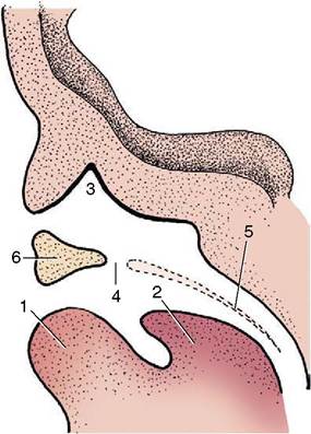
Figure 3-57 Sagittal section through the nasal and oral cavity of a young embryo. 1, Lower lip; 2, tongue; 3, nasal cavity; 4, primitive choana (future incisive duct); 5, position of future secondary palate; 6, primary palate.
edge of the septum between the nasal fossae; a horizontal shelf is thus formed between the nasal fossae and the mouth. Fusion of the residual primary palate (the region of the incisive papilla) with the palatine processes is almost complete but leaves open the small passages that become the incisive ducts. The shelf that now divides the nasal and oral cavities constitutes the secondary (definitive) palate, which later differentiates into rostral hard and caudal soft parts. The mechanism of its formation is not wholly understood; the timing is critical because the stage at which the secondary palate forms is normally soon followed by a marked widening
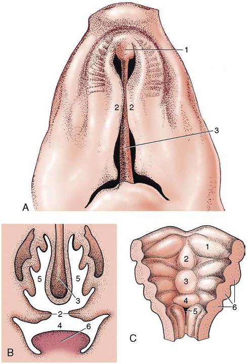
Figure 3-58 A, Ventral view of the development of the palate (pig).
B, Transverse section through oral and nasal cavity before closure of the secondary palate. 1, primary palate; 2, palatine processes (secondary palate); 3, nasal septum; 4, oral cavity; 5, nasal cavity; 6, tongue. C, Development of the tongue in the floor of the oral cavity. 1, Distal (lateral) tongue swelling; 2, median tongue swelling; 3, proximal tongue swelling; 4, primordium of epiglottis; 5, laryngeal entrance; 6, pharyngeal arches.of the head. If reorientation of the palatine processes is delayed, they are too short to bridge the gap and fail to fuse with each other and with the ventral edge of the nasal septum, which leaves the secondary palate divided by a median fissure through which the nasal and oral cavities communicate. The consequences of this anomaly (cleft palate) can be severe, not least because of resulting difficulties in feeding from the teat.
The division of the mouth cavity into its vestibular and central parts is foreshadowed by the appearance of ectodermal thickenings that run parallel to the margins of both the maxillary and the mandibular processes. These thickenings are soon transformed into grooves, known as labiogingival grooves, as they mark the division of the lips from the outer aspect of the gums; deepening of the grooves creates and then enlarges the vestibular space. A second, similar formation internal to the labiogingival groove of the mandibular process separates the gum from the tongue now developing in the floor of the mouth.
The salivary glands, both major and minor, are formed from solid outgrowths of epithelium that push into the underlying mesenchyme. These branch repeatedly and become canalized to form both gland acini and ducts. It is tempting to suppose that their sites of origin correspond with the points of entry of the adult ducts; however, some evidence suggests that the openings may be relocated when grooves in the oral epithelium are bridged over, extending the ducts.
The tongue develops in the floor of the mouth.
It has a complicated origin, being formed by the mergence of several swellings (see Figure 3-58, C). One, a median (distal) tongue swelling, appears on the pharyngeal floor between the lower ends of the mandibular arches and later fuses with more lateral swellings that appear over the adjacent parts of these arches. A more caudal (proximal) swelling extends from the floor onto the ventral parts of the second, third, and, possibly, fourth pharyngeal arches. The caudal swelling divides as follows: the caudal part becomes the epiglottis and the rostral part blends with the other contributions to the tongue. The thyroid gland develops from the pharyngeal floor between the median and proximal swellings. The substance of the tongue is supposed to derive mainly from myotomes of occipital somites. It is alleged that material from these myotomes migrates forward under the floor of the mouth, and although the evidence is not wholly convincing, the theory satisfactorily accounts for the innervation of the lingual muscles by the hypoglossal nerve, which is the nerve specific to the occipital somites. The sensory supply to the lingual epithelium involves the mandibular, fascial, glossopharyngeal, and vagus nerves, which are the nerves associated with the first, second, third, and fourth arches.The separation of the tongue from the floor is gradual; it is more complete for the part that forms the body than for that that forms the root.
The first indications of the teeth are ribbonlike thickenings of epithelium internal to the labiogingival thickenings. The thickenings extend as plates, dental laminae, into the subjacent mesenchyme (Figure 3-59); quite soon a linear series of knoblike swellings buds from the deep margin of each. The swellings represent the enamel organs of the temporary teeth, and their number corresponds to the dental formula of the species. Occasionally it is greater; the disparity occurs when primordia appear (and possibly develop quite far) for teeth that later regress without erupting.
The upper incisors of ruminants are examples of teeth whose development is aborted in this way.The mesenchyme condenses against the free surfaces of each bud; when the bud shortly invaginates, the mesenchyme now known as the dental papilla fills the resulting cup. The whole tooth germ, the enamel organ together with the dental papilla, is enclosed by a mesenchymal thickening that merges with the papilla at its base, forming the dental sac or follicle.
The enamel organ consists of an inner epithelium (over the concave surface applied to the dental papilla), an outer epithelium (over the convex surface facing the dental follicle), and an intervening sparsely cellular tissue (enamel reticulum) (see Figure 3-59). The cells of the inner dental epithelium are known as ameloblasts because they produce enamel. Enamel formation begins over the center of the crown but soon spreads outward from this focus. As the layer thickens, the ameloblasts retreat in a centrifugal direction until finally they meet and fuse with the outer dental epithelium to form an epithelial cuticle over the crown.
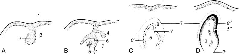
Figure 3-59 A, Development of dental plate. B, Development of an enamel organ. C, Enamel organ. D, Deciduous tooth before eruption. 1, Epithelium of oral cavity; 2, dental plate; 3, mesenchyme; 4, bud of a permanent tooth; 5, dental papilla; 5', odontoblasts (differentiated from the outer cell layer of the papilla); 5", dentine; 6, inner dental epithelium (future ameloblasts); 6, ameloblasts; 6", enamel; 7, outer dental epithelium; 7', transition of inner and outer dental epithelia (where root formation occurs); 8, enamel reticulum.
Meanwhile, certain cells of the mesodermal papilla have become arranged in a sheet facing the ameloblasts. Because they produce dentine, they are known as odontoblasts. The first dentine also appears toward the center of the crown, a little later than the first deposition of enamel. Thereafter dentine deposition also spreads out in all directions. As the layer thickens the odontoblasts withdraw in a centripetal direction, and when dentine production has ceased, they remain as a covering to the pulp, which is the surviving less differentiated portion of the original papilla.
The root of the tooth is initially ensheathed by a prolongation of the enamel organ not producing enamel. The sheath later breaks down when the follicular tissue produces cement to encase the dentine of the root.
After the enamel organs of the temporary teeth have appeared the dental lamina undergoes extensive destruction. However, its free edge remains to produce a second crop of buds, the enamel organs of the replacement teeth; these remain dormant until activated to replicate the sequence that created the temporary teeth.