THE MOUTH
The wide gape of carnivores is made possible by the caudal situation of the angles of the mouth and the correspondingly short cheeks. The interior of the mouth, including the oropharynx, is therefore easily examined.
The edge of the lower lip carries blunt papillae. The upper lip is pendulous and presses on the lower one, which is everted near the commissure in certain breeds with ample head skin, such as the Spaniel (Figure 11-6, A, and Figure 11-13). The resulting folds predispose to infection. The general looseness of the lips creates a large vestibule—an advantage when administering liquid medicines, which then escape behind the cheek teeth into the central cavity.The ducts of the parotid (Figures 11-6, AJ13-BH3 and 11-14/3) and zygomatic (Figures 11-10/3 and 1127/8) salivary glands open into the vestibule: the former by a single orifice in a small papilla opposite the upper
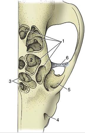
Figure 11-11 The canine frontal sinuses, dorsal view. 1, Lateral frontal sinus; 2, ethmoidal concha invading the sinus; 3, medial and rostral frontal sinuses; 4, infraorbital foramen; 5, orbit; 6, orbital ligament.
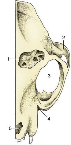
Figure 11-12 The feline frontal sinus, dorsal view. 1,
Frontal sinus, opened; 2, zygomatic arch; 3, orbit; 4, position of infraorbital foramen; 5, nasal aperture.
fourth premolar P4, and the latter by a row of four or five orifices on a mucosal ridge a little farther caudally. The ducts of the mandibular and compact (monosto- matic) sublingual glands open to the floor of the mouth at the sublingual caruncle. They run below the mucous
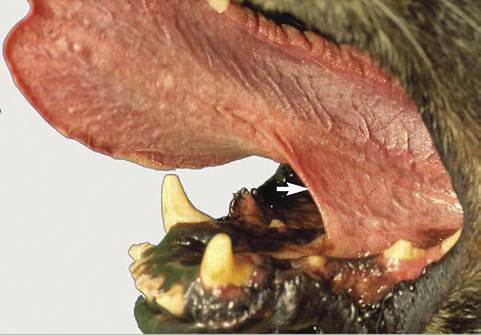
Figure 11-13 Tongue with frenulum.
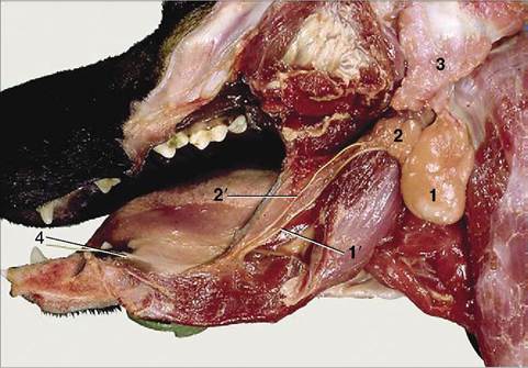
Figure 11-14 Salivary glands. 1, Mandibular gland; 1', mandibular duct; 2, sublingual gland, monostomatic part; 2', its duct; 3, parotid gland; 4, sublingual caruncle.
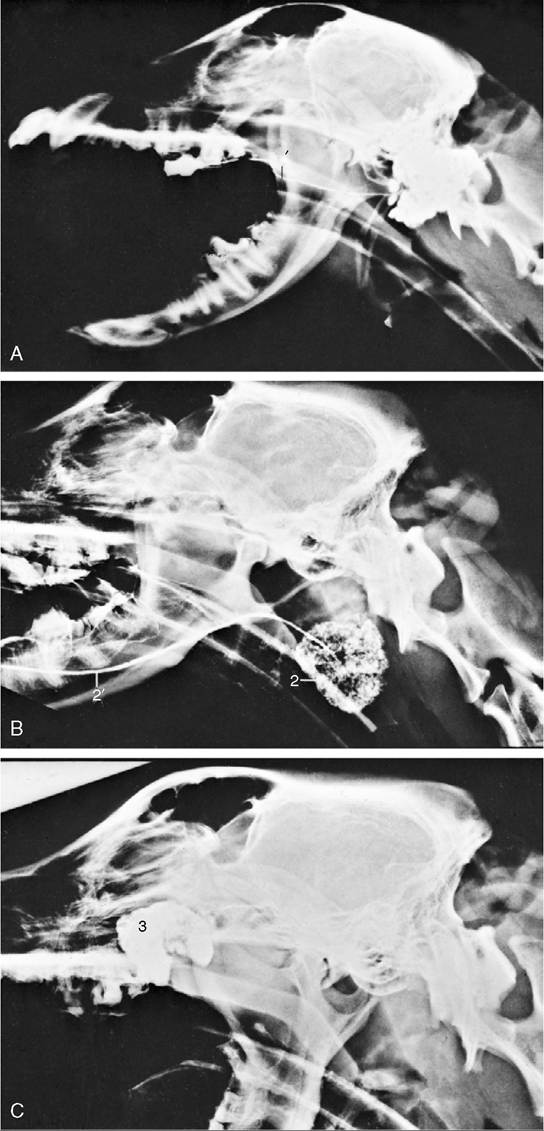
membrane that connects the side of the tongue with the gums; when a duct is damaged, saliva may escape to form a large mucosal swelling (ranula) lateral to the tongue. The larger salivary ducts are occasionally cannulated to remove obstructions or to inject a contrast medium for radiographic examination (sialography, Figure 11-15).
The oral cavity proper, like the nasal cavity above, widens from front to back before contracting at the level of the palatoglossal arches, beyond which it is continued by the oropharynx.
The hard palate presents transverse ridges and a prominent incisive papilla (see Figure 3-5). The slit to each side of the incisive papilla opens into an incisive duct that extends caudodorsally for 1 or 2 cm through the palatine fissure to open onto the floor of the nasal cavity. Before doing so, the duct communicates with the cavity of the vomeronasal organ. The Flehmen reaction
1
1
Figure 11-15 Contrast medium outlining the canine parotid (A), mandibular (B), and zygomatic (C) glands. 1, Parotid gland; 1', duct; 2, mandibular gland; 2', duct; 3, zygomatic gland.

Figure 11-16 Tomcat demonstrating Flehmen.
associated with the perception of pheromones is exhibited in both dogs and cats but is less clearly demonstrated than in animals such as in horses (Figure 11-16).
The oral mucosa, generally pink, may be pigmented locally. The wide and flat apex of the tongue is depressed centrally (like a spoon) when liquids are lapped. A short median rod (lyssa) of connective, muscular, and cartilaginous tissue is embedded close to the ventral surface of the tongue. Its significance is not known, although a fanciful connection with rabies was postulated in former times.
The dorsal surface of the tongue is roughened by papillae. Filiform papillae predominate but are replaced by stouter conical papillae toward the root; both have protective and mechanical functions. Other papillae are concerned with the perception of taste; round fungiform papillae are dotted among the filiform papillae; foliate papillae, represented by a few shallow grooves, are present on the lateral border, near the palatoglossal arch; and four to six vallate papillae form a rostrally open V on the root (Figure 11-17). The tongue of the newborn is fringed with lacelike (marginal) papillae that persist for the first 2 weeks and are thought to assist in fitting the tongue to the dam’s teat.
The oral cavity of the cat is short and wide and is easily examined in cooperative subjects (Figure 11-18). The abrasive nature of the cat’s tongue is due to the strong keratinization of the epithelium of the large conical papillae that replace the delicate filiform papillae of most species. On the dorsum of the tongue these papillae are caudally directed and hooked; this assists in grooming but makes it more difficult to eject threadlike objects that have been taken into the mouth (Figure 11-19 and Figure 11-20). Hairs removed from the coat during grooming therefore accumulate in the stomach
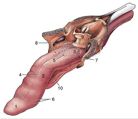
Figure 11-17 The tongue of the dog. The soft palate and the esophagus are sectioned in the median plane. 1, Apex; 2, body; 3, root, forming floor of oropharynx; 4, median groove; 5, vallate papilla; 6, fungiform papillae; 7, palatoglossal arch; 8, palatine tonsil in tonsillar fossa; 9, epiglottis; 10, frenulum.
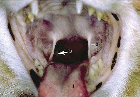
Figure. 11-18 Oropharynx (cat). 1, Tongue; 2, palatoglossal arch; 3, position of right palatine tonsil (arrow).
(hairballs); they mingle with the ingesta and may be expelled with the feces or ejected through the mouth.
In addition to diffuse labial salivary glands, the lips of cats contain large sebaceous and apocrine glands. The secretion of these circumoral glands is used in grooming and may be frequently rubbed off on objects, apparently as a scent marker substance (see Figure 10-11).
Congenital clefts of the primary (harelip) or secondary palate has been reported in both dogs and cats,
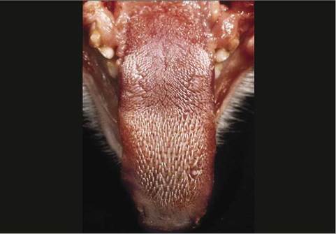
Figure 11-19 Tongue (cat) with papillae.
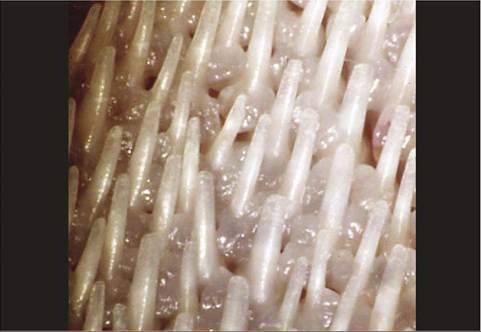
Figure 11-20 Enlargement showing caudally directed keratinized filiform papillae (cat).
especially Siamese. In dogs, the incidence of cleft palate is higher in brachycephalic breeds, although other breeds (Labrador, Cocker Spaniel) may be affected. The primary palate forms the lips and premaxilla, and the secondary palate forms the hard and soft palates. The incomplete closure of these structures is attributed to inherited and environmental factors. Clefts may be inherited as either recessive or irregular dominant traits. Toxic agents and intrauterine viral infections can produce animals with clefts if the insult occurs at a very specific time in fetal development (25th to 28th day in dogs).
Fractures of the mandible and separation at the symphysis are fairly common in both species, often as the result of traffic accidents. Concurrent involvement of the maxilla, nasal structures, teeth, and soft tissues of the face is more frequent in cats that have fallen from heights.