THE PARANASAL SINUSES
The sinus system of the dog is poorly developed. The largest sinus is the frontal one, which occupies much of the frontal bone including its zygomatic process, which is separated from its fellow by a median septum.
It may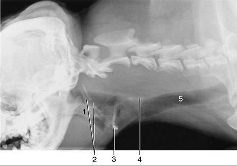
Figure 11-9 Radiograph of the cramped pharyngeal region of the brachiocephalic dog. (The space available is rather cramped.) 1, Soft palate; 2, hyoid apparatus; 3, basihyoid; 4, cricoid cartilage; 5, trachea.
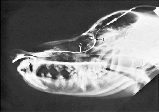
Figure 11-8 Contrast medium outlining the canine nasolacrimal duct in a radiograph. 1, Position of ventral punctum; 2, nasolacrimal duct; 3, opening of duct at the nostril.
extend to the level of the temporomandibular joints in larger animals (especially if long-headed) (Figure 11-11). Each sinus is composed of three cavities (lateral, medial, and rostral), which communicate separately with the nasal cavity via nasofrontal openings (ethmoidal meatuses). The lateral compartment is the largest and may be subdivided by incomplete septa; ethmoturbinates are present in its rostral part. The medial and rostral compartments are also filled with ethmoturbinates, which hampers their identification on radiographs. The ethmoturbinates are covered with olfactory mucosa, in contrast to the sinus walls that are lined with nonolfactory mucoperiosteum. The sinuses are smaller and may even be absent in brachycephalic breeds. Absence is not associated with clinical signs and is usually only found when radiographs are made for another reason.
The sinus system of the cat comprises frontal, sphenoidal, and maxillary compartments, among which the frontal is the most important (Figure 11-7, B, and Figure 11-12/7). It occupies the same general position as the corresponding sinus in the dog, but it is undivided and extends rather far ventrally within the medial wall of the orbit.
The communication with the nasal cavity is in its rostral part and may provide ineffective drainage in the bacterial sinusitis that commonly complicates viral infections of the upper respiratory tract. Surgical drainage may then be required. In mature cats, the sinus can be surgically approached just lateral to the midline, on the line connecting the rostral margins of the supraorbital processes. In 3 to 4 month-old kittens the approach is made midway between the line connecting the rostral margins of the supraorbital processes and that connecting the medial angles of the eyes.In both dogs and cats, the maxillary sinus (Figure 11-10/73) communicates so freely with the nasal cavity that the term nasal recess is preferred. It is not a true sinus because it is not formed between two plates of maxillary bone but is bounded by the maxilla laterally and the ethmoid medially. The recess occupies the face immediately rostral to the orbit, above the roots of the last three cheek teeth, and communicates with the middle meatus by a wide nasomaxillary opening flanked by the nasal conchae. The recess houses on its lateral wall the broad, flat, lateral nasal gland, which appears as a thickening of the mucosa. Root abscesses of the sectorial tooth P4 may break into the recess and later
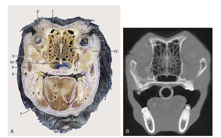
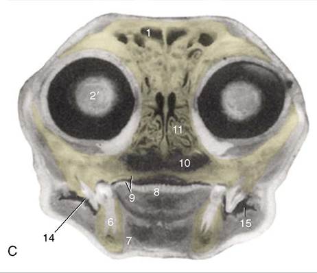
Figure 11-10 Transverse section of the canine (A) and feline (C) heads through the rostral part of the orbit, rostral surface. B, CT scan (bone window) of canine head at the level of A. 1, Frontal sinus; 2, orbital structures; 2', eye; 3, zygomatic gland; 4, masseter; 5, facial vein; 6, mandible; 7, digastricus; 8, tongue; 9, oral cavity and hard palate; 10, choana; 11, ethmoidal conchae; 12, zygomatic arch; 13, maxillary recess; 14, sectorial teeth, P4 engaging M1; 15, oral vestibule.
onto the surface of the skull. Surgical drainage is most conveniently achieved by the extraction of the sectorial tooth to open a passage to the mouth; the presence of the infraorbital canal makes the direct lateral approach unwise.
In cats, a small sphenoidal sinus is present; the similar cavity found in dogs is filled with ethmoturbinates.