» The Mouth and Dentition
The animal's inability to open its mouth widely and problems with restraint make it difficult to examine the long and narrow mouth of the conscious animal. The ridges of the roof of the rostral part of the cavity end abruptly at the boundary of the soft palate, where the two discrete tonsils of the soft palate, which correspond to the tonsils embedded in the lateral walls of the oropharynx of other species, are found.
These tonsils are cut in routine meat inspection.The pointed tongue occupies the floor. In the newborn, the tongue is fringed with lacelike marginal papillae (Fig. 32.7/3), which persist for the first 2 or 3 weeks of life; because they swell visibly preparatory to contact with the teat, they are believed to help seal the mouth about the teat when sucking.
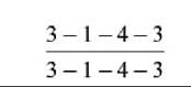
Pigs have the most complete dentition of any domestic animal (see Fig. 3.18); the formula for the permanent dentition is:
The straight lower incisors meet the curved upper incisors to provide a potential grasping action (Fig. 32.8). The curved canine teeth, or tusks, are firmly embedded in the jaws. In boars the roots remain open, and the tusks grow throughout life, providing these animals with formidable weapons; however, in sows growth ceases after 2 years, and their smaller tusks do not project from the mouth. The tusks of boars are often cut short, sometimes without benefit of anesthesia. The crowns of the check teeth increase in both length and breadth from first to last in the series. The occlusal surfaces of the molars show many irregularities and are ideally adapted for crushing food.
FIG. 32.3 Head, superficial dissection. 1, Cut fasciculi of levator nasolabialis; 2, caninus; 3, levator labii superioris; 4, malaris; 5, facial vein; 6, dorsal nasal vein; 7, frontal vein; 8, levator anguli oculi; 9, frontoscutularis; 10, lateral retropharyngeal lymph node; 11, parotidoauricularis; 12, trapezius; 13, cleidooccipitalis; 14, omotransversarius; 15, parotid gland; 16, sternocephalicus; 17, sternohyoideus; 18, parotid duct; 19 and 20, ventral and dorsal buccal branches of facial nerve, respectively; 21, transverse facial nerve; 22, inferior labial vein; 23, superior labial vein; 24, masseter; 25, mental hairs and gland; 26, depressor labii inferioris; 27, mentalis; 28, depressor labii superioris; 29, orbicularis oris; 30, mandible.
FIG. 32.4 Paramedian section of the skull. 1, Dorsal turbinate bone, fenestrated at 6 to show conchal sinus; 2, ventral turbinate bone; 3, hard palate; 4, choana; 5, ethmoturbinates in fundus of nasal cavity; 6, conchal sinus; 7, portion of frontal sinus exposed by paramedian saw cut; 8, position of orbit; 9, cranial cavity; 10, optic canal; 11, petrous temporal bone; 12, fossa for hypophysis; 13, sphenoid sinus; 14, tympanic bulla; 15, paracondylar process; 16, hamulus of pterygoid bone. I3, third upper incisor; M1, first upper molar; P1, first upper premolar.
FIG. 32.5 Transverse sections of the nose of piglets treated with the toxin causing atrophic rhinitis. (A) The piglet is treated with a low dose. (B) The piglet is treated with an activated dose. (C) The piglet is treated with an inactivated dose.
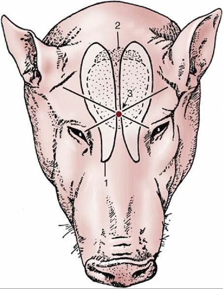
FIG. 32.6 Head of a 9-month-old pig. 1, Outline of frontal sinuses; 2, position of brain; 3, point at which pig is best shot for stunning at slaughter.
Table 32.1 summarizes the ages at which different teeth erupt and are replaced. The deciduous incisors and canines with which the piglet is born are known as needle teeth. They project laterally from the gums and, being very sharp, may injure the mother's teat or any littermate in competition for this. They are therefore commonly nipped off within hours of birth; the procedure requires some care if the marginal lingual papillae are not to be injured. The dentition is normally complete by the age of 18 months, long after sexual maturity is reached.
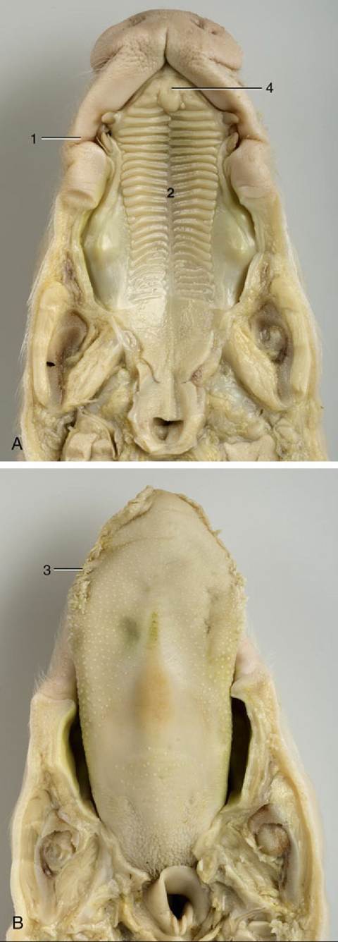
FIG. 32.7 (A) The roof and (B) the floor of the mouth of a newborn piglet.
1, Permanent notch in upper lip opposite tusk; 2, hard palate with ridges; 3, lingual marginal papillae; 4, incisive papilla.The large parotid gland lies ventral to the base of the ear (Fig. 32.3/15). It extends only a little way over the masseter muscle rostrally, but its cervical angle reaches beyond the middle of the neck under cover of the cutaneous muscle. It has numerous relations to the structures within the visceral space of the neck. Its duct crosses the mandibular gland and curves around the ventral border of the mandible to gain the face and open into the buccal cavity. The smaller rounded mandibular gland lies partly medial to the mandible and partly deep to the parotid. Its duct runs alongside the sublingual gland to open at the sublingual caruncle. Both parts of the sublingual gland are present; they drain in the usual way.
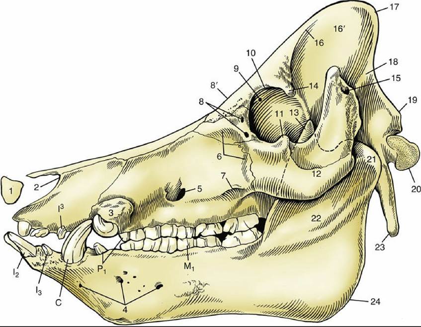
FIG. 32.8 Skull of a boar. 1, Rostral bone; 2, nasoincisive notch; 3, canine eminence; 4, lateral mental foramina; 5, infraorbital foramen; 6, fossa canina; 7, facial crest; 8, lacrimal foramina; 8', location of supraorbital foramen on dorsal surface; 9, orbital end of supraorbital canal; 10, orbital rim; 11, frontal process of zygomatic bone; 12, zygomatic arch; 13, coronoid process of mandible; 14, zygomatic process of frontal bone; 15, external acoustic meatus; 16, temporal line; 16', temporal fossa; 17, nuchal crest; 18, temporal crest; 19, nuchal tubercle; 20, occipital condyle; 21, condylar process of mandible; 22, ramus of mandible; 23, paracondylar process; 24, angle of mandible; C, canine teeth (tusks); I2,13, and I3, first and second lower and third upper incisors; M1, first lower molar; P1, first premolars.
» TABLE 32.1
Eruption Dates of Porcine Teeth
| Temporary Tooth | Permanent Tooth | |
| Incisor 1 | 1-3 weeks | 11-18 months |
| Incisor 2 | 8-12 weeks | 14-18 months |
| Incisor 3 | Before birth | 8-12 months |
| Canine | Before birth | 8-12 months |
| Premolar 1 | 4-8 months | — |
| Premolar 2 | 6-12 weeks | 12-16 months |
| Premolar 3 | 1-3 weeks | 12-16 months |
| Premolar 4 | 2-5 weeks | 12-16 months |
| Molar 1 | — | 4-8 months |
| Molar 2 | — | 7-13 months |
| Molar 3 | — | 17-22 months |