THE MUSCLES OF FACIAL EXPRESSION
Many clinically important features are revealed as soon as the skin is removed. Large areas of the skull are not covered by any considerable thickness of soft tissue and are therefore vulnerable to injury.
These areas include the dorsal aspect of the nose, the forehead, and part of the temple, in addition to much of the mandible. Prominent landmarks include the facial crest, which runs parallel to the dorsum of the nose; it begins above the rostral margin of the fourth cheek tooth, continues into the zygomatic arch, which forms the lower margin of the orbit, and extends to the temporomandibular joint (Figure 18-6Z√). The joint itself is easily located by the salience of the lateral aspect of the condyle, directly before the palpable caudal margin of the mandible. The identification becomes more certain if the animal can be induced to perform chewing movements. The ventral margin of the mandible is also prominent, particularly the half that lies rostral to the masseter muscle. A shallow notch in the bone directly in front of the muscle conveys the facial vessels and parotid duct from the intermandibular space to the face.The incomplete sheet of cutaneous muscle over the lateral aspect of the head is best developed where it merges with the orbicularis oris around the opening of the mouth.
A few individual mimetic muscles deserve notice. The levator labii superioris arises over the maxilla and runs dorsorostrally to form a common tendon with its fellow of the other side (Figure 18-7Z7); the tendon, which is enclosed within a synovial sheath, descends between the nostrils to splay out within the upper lip. This muscle is responsible for the lip curl (Flehmen) seen in certain circumstances, including sexual excitement. The levator belly is easily palpated, and because it covers the infraorbital foramen, it must be pushed dorsally to locate the emergent infraorbital nerve.
This foramen lies along the line joining the nasoincisive notch to the rostral end of the facial crest.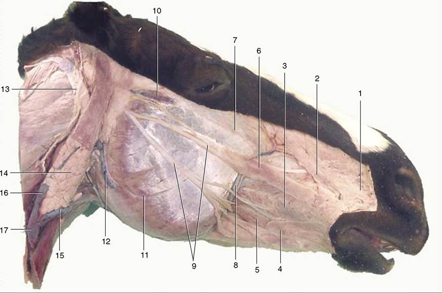
Figure 18-7 Superficial dissection of the head. 1, Caninus; 2, levator nasolabialis; 3, buccinator; 4, stump of cutaneous muscle joining orbicularis oris; 5, depressor labii inferioris; 6, zygomaticus; 7, levator labii superioris; 8, facial artery and vein; 9, buccal branches of facial nerve; 10, transverse facial artery and vein and transverse facial branch of auriculotemporal nerve; 11, masseter; 12, masseteric artery and vein; 13, great auricular nerve (C2); 14, parotid gland; 15, linguofacial vein; 16, maxillary vein; 17, external jugular vein.
The depressor labii inferioris (Figure 18-7/5) arises with the buccinator from the alveolar margin and adjacent part of the mandible under cover of the masseter. It can be identified as a rounded cord running rostrally over the body of the bone. The tendon covers the mental foramen, located about 2 to 3 cm caudal to the angle of the mouth, and this is readily palpable when the muscle is slid aside. The buccinator (Figure 18-7/5) has a well-marked herring-bone structure and is partly covered by the masseter. It is important in returning food to the central cavity of the mouth, preventing its accumulation in the oral vestibule.