THE MUSCLES OF THE FORELIMB
The muscles of the forelimb comprise the girdle musculature, passing between the trunk and the limb, and the intrinsic musculature.
Girdle Muscles
The girdle muscles join the forelimb to the trunk, forming a connection known as a synsarcosis that substitutes for a conventional joint.
When the animal is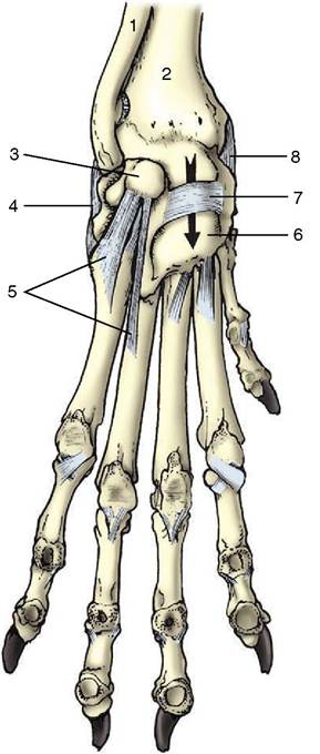
Figure 2-53 Left carpal joint of the dog, palmar view. 1, Ulna; 2, radius; 3, accessory carpal; 4, lateral collateral ligament; 5, distal ligaments of accessory carpal; 6, palmar carpal ligament; 7, flexor retinaculum; 8, medial collateral ligament; the arrow is in the carpal canal.
standing, some of the muscles of the synsarcosis (the serratus ventralis and pectoralis profundus) sling the body between the forelimbs to which they transmit the weight of the head, neck, and cranial part of the trunk (Figure 2-54). These and other girdle muscles can also stabilize the scapula against external forces, preventing its displacement or rotation. A good example of this role is supplied by a cat pouncing on a mouse or plaything with forelimbs rigidly braced against the trunk. During progression the same muscles resolve into antagonistic groups that control the swing of the limb; one group advances (protracts) the limb, the other retracts it. For these actions to be understood, it is necessary to appreciate that the scapula may be moved against the chest wall in two different ways. In one, the bone is rotated about a transverse axis located toward its upper end. The position of this axis, which is of course imaginary, is fixed by the balance of opposing muscles, chiefly the rhomboideus and serratus ventralis,
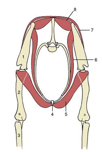
Figure 2-54 Muscular suspension of the thorax between the forelimbs (dog).
1, Scapula; 2, humerus; 3, radius and ulna; 4, sternum; 5, pectoralis profundus (ascendens); 6, serratus ventralis; 7, trapezius; 8, rhomboideus.which both attach on the dorsal part of the scapula. In the other movement, the whole bone is shifted on the thoracic wall. It is slid downward and forward as the limb is advanced and upward and backward in recovery during retraction. This movement of the scapula, which adds usefully to the length of the stride, is permitted by the looseness of the connective tissue that intervenes between the limb and the trunk where there exists a potential space, the axilla, corresponding to the human armpit. The axilla also gives passage to the nerves and vessels entering the limb from the trunk, and it contains the axillary lymph nodes.
For the purpose of description, the girdle muscles can be considered in two layers.
The Superficial Layer. This consists of a cranial group supplied mostly by the accessory nerve, the latissimus dorsi more caudally, and the two superficial pectoral muscles ventrally. The cranial group comprises the trapezius, omotransversarius, and brachiocephalicus.
The trapezius (Figure 2-55Z5,5,) is thin. It takes origin from the middorsal raphe and supraspinous ligament, extending from about the level of the second cervical to that of the ninth thoracic vertebra, and converges to insert on the spine of the scapula. It consists of two fleshy parts, cervical and thoracic, usually separated by an intermediate aponeurosis. The fibers of the cervical part run caudoventrally to attach along the
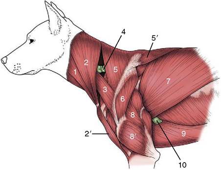
Figure 2-55 Superficial muscles of the shoulder and arm. 1, Sternocephalicus; 2, 2', brachiocephalicus: cleidocervicalis and cleidobrachialis; 3, omotransversarius; 4, superficial cervical lymph node; 5, 5', cervical and thoracic parts of trapezius; 6, deltoideus; 7, latissimus dorsi; 8, 8', long and lateral heads of triceps; 9, pectoralis profundus (ascendens); 10, accessory axillary lymph node.
greater part of the length of the scapular spine; those of the thoracic part run cranioventrally to a more confined insertion on the tuberous thickening of the spine. The trapezius may raise the scapula against the trunk and swing the ventral angle of the bone cranially, thus advancing the limb.
The omotransversarius (Figure 2-55Z3) is a narrow muscle that extends between the transverse processes of the atlas (and possibly also the succeeding vertebrae) and the acromion and adjacent part of the scapula. It assists in advancement of the limb.
The brachiocephalicus (Figure 2-55Z2,2,) is more complex, being formed by the union of two elements that are separated by the clavicle in less specialized mammals. In these the caudal part (cleidobrachialis) passes between the clavicle and the humerus and is a component of the deltoideus muscle. The cranial part passes cranially from the clavicle to several attachments in the head and neck. These attachments vary among species and hence a rather bewildering array of names for particular units exists: cleidooccipitalis, cleidomas- toideus, and so forth. In domestic species the two parts join in tandem, and the clavicle is generally reduced to a fibrous intersection in the combined muscle at the level of the shoulder joint, although vestigial ossifications are present in the dog and cat. Brachiocephalicus is a most appropriate name for the whole complex since it does not specify precise attachments. The brachiocephalicus advances the limb, possibly also extending the shoulder joint, when the cranial attachment is fixed and the limb is free to move; in contrast, when the limb is fixed and the head is free, it draws the head and neck ventrally when acting bilaterally and toward the side when acting unilaterally.
The muscles supplied by the accessory nerve split from a single primordium in the embryo. However, the caudal part of the brachiocephalicus of deltoid origin retains the appropriate innervation by the axillary nerve.
The latissimus dorsi (Figure 2-55/7) has a very broad origin from the thoracolumbar fascia and converges to an insertion on the teres tuberosity of the humerus. The most cranial fibers, which are also the most vertical, cover the caudal angle of the scapula and strap it against the chest. The muscle retracts the free limb and may also flex the shoulder joint. On the other hand, when the limb is advanced and the foot firmly planted on the ground, the latissimus may draw the trunk forward. It may be regarded as antagonist to the brachiocephalicus. It is supplied by a local branch (thoracodorsal nerve) of the brachial plexus.
Two superficial pectoral muscles (Figure 2-41/5,6) arise, one behind the other, from the cranial part of the sternum. The cranial muscle (pectoralis descendens) terminates on the crest of the humerus, distal to the deltoid tuberosity. The caudal muscle (pectoralis transversus) descends over the medial aspect of the arm and in the larger species continues distally over the elbow joint, covering the median artery and nerve, to insert into the medial fascia of the forearm. Both muscles adduct the limb, which is an action that may be understood to embrace the sideways shift of the trunk toward a previously abducted limb. It seems probable that they may also assist protraction or retraction, depending on the initial position of the limb relative to the trunk. They are supplied by local branches (cranial pectoral nerves) from the brachial plexus.
The Deep Layer. This comprises the rhomboideus dorsally, the serratus ventralis medially, and the pectoralis profundus ventrally.
The rhomboideus (Figure 2-54/8) takes origin from median connective tissue structures extending from the poll to the withers and lies deep to the trapezius. It always presents cervical and thoracic parts and in carnivores has an additional, capital, part. All attach to the dorsal border and adjacent area on the medial surface of the scapula. Although the fiber courses differ in their relation to the axis of rotation of the scapula, most seem able to draw the dorsal part of the bone cranially, thereby retracting the limb.
The muscle may also raise the limb and hold it firmly against the trunk. It is supplied from the brachial plexus in the dog, but in some species it is also supplied by dorsal branches of local spinal nerves, which is unusual for a limb muscle.The serratus ventralis (Figure 2-54/6) is a large fanshaped muscle that takes an extensive origin by separate digitations from the fourth cervical vertebra to the tenth rib. The fibers run dorsally to terminate on a well- defined area on the medial aspect of the scapula and scapular cartilage. The direction of the fibers indicates that this muscle must play a large part in supporting the weight of the trunk, and in the larger species it is better adapted to this function by the presence of a strong fascial covering and intersections. The cervical portion of the muscle, which inserts craniodorsal to the axis of scapular rotation, can retract the limb; the caudal portion, which inserts caudodorsal to the axis, can advance the limb. When acting unilaterally, the cervical fibers may also draw the neck to that side; when acting bilaterally, they raise the neck. The thoracic part is a potential inspiratory muscle, although it is not normally used in that capacity. The innervation is mainly by a branch (long thoracic nerve) of the brachial plexus.
The pectoralis profundus (Figure 2-55/9) may be considered as having cranial and caudal parts. The cranial part, well formed only in the horse and pig, probably corresponds to the subclavius of other mammals and is now so named officially. Both parts (or muscles) arise from the ventral aspect of the length of the sternum and adjacent cartilages, and the most caudal fibers extend beyond this onto the abdominal floor. In the horse and pig the subclavius passes dorsally along the leading edge of the scapula, attaching to the supraspinatus (see Figure 23-5, A/2). The larger caudal part, also known as the pectoralis ascendens, inserts on the lesser tubercle of the humerus. Both play a role, secondary to that of the serratus ventralis, in slinging the trunk between the forelimbs.
They may also act as retractors of the forelimb when this is free. When the limb is advanced and fixed, they draw the trunk forward, toward the limb. The nerves are local branches (caudal pectoral nerves) of the brachial plexus.Intrinsic Muscles of the Forelimb
The intrinsic muscles are conveniently grouped by their common location, actions, and innervations.
Muscles Acting Primarily on the Shoulder Joint. The muscles acting on the shoulder joint are arranged in lateral, medial, and caudal groups.
The lateral group comprises the supraspinatus and infraspinatus, which arise from and fill the corresponding fossae of the scapula. The supraspinatus (Figure 2-56/3) terminates on the summits of both tubercles of the humerus. The infraspinatus inserts by a tendon that splits into a shorter deep part, which attaches to the summit, and a longer superficial part, which attaches to the lateral face of the (caudal part of the) greater tubercle; a bursa between the bone and the longer tendon may be the seat of a painful inflammation. Both muscles brace the joint laterally. The supraspinatus tendon passes cranial to the axis of rotation, and it may therefore also extend the shoulder. It is sometimes asserted
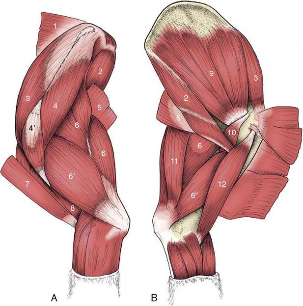
Figure 2-56 Intrinsic muscles of the left shoulder and arm of the dog, lateral (A) and medial (B) views. 1, Rhomboideus; 2, teres major; 3, supraspinatus; 4, 4', scapular and acromial parts of deltoideus; 5, latissimus dorsi; 6, 6', 6", long, lateral, and medial heads of triceps; 7, brachiocephalicus; 8, brachialis; 9, subscapularis; 10, coracobrachialis; 11, tensor fasciae antebrachii; 12, biceps.
that the infraspinatus tendon passes cranial or caudal to the axis of rotation depending on the actual position of the joint and may then further extend the already extended joint or further flex the already flexed joint; clearly, it is unlikely to be very effective in either role. Both muscles are supplied by the suprascapular nerve from the brachial plexus.
The medial group comprises the subscapularis and coracobrachialis. The subscapularis (Figure 2—56/9) arises over much of the deep surface of the scapula and inserts on the medial tubercle of the humerus, distal to the axis of the shoulder joint. It braces the medial aspect of the joint. It is also a potential adductor of the arm and, like the infraspinatus, has an equivocal relationship to flexion and extension of the shoulder. It is supplied by the subscapular nerve from the brachial plexus. The coracobrachialis (Figure 2—56/10) extends between the medial aspect of the supraglenoid tubercle and the proximal part of the shaft of the humerus. Too small to be of real significance, it is a fixator of the shoulder with the same equivocal relationship to shoulder flexion and extension. It is supplied by the proximal branch of the musculocutaneous nerve from the brachial plexus.
The caudal or flexor group comprises the deltoideus, teres major, and teres minor. The deltoideus has one head of origin in the horse and two in species possessing an acromion (Figure 2-56/4,4'). The constant head arises from the caudal border and spine of the scapula; the inconstant second head arises from the acromion. Both insert on the deltoid tuberosity of the humerus. The teres major (Figure 2-56/2) arises from the dorsal part of the caudal margin of the scapula and terminates on the teres tuberosity, midway down the humerus. The relatively insignificant teres minor lies over the caudola- teral aspect of the joint between the deltoideus and infraspinatus. These three muscles are clearly primarily flexor; the deltoideus may also be an abductor and an outward rotator of the arm. The group is supplied by the axillary nerve from the brachial plexus.
In contrast to the well-defined group of flexors, it seems that no muscles are clearly established as primarily extensors of the shoulder. The potential candidates, brachiocephalicus, biceps brachii, supraspinatus, and pectoralis ascendens, have other, apparently more important roles.
Muscles Acting Primarily on the Elbow Joint. There are extensor and flexor groups. The extensor group, which largely fills the angle between the scapula and humerus, consists of the triceps brachii, tensor fasciae antebrachii, and anconeus. The large and powerful triceps brachii (Figure 2—56/6,6',6") possesses three heads of origin (four in the dog). The long head, which arises from the caudal margin of the scapula, is potentially also a flexor of the shoulder. The lateral, medial, and (in the dog) accessory head(s) arise from the shaft of the humerus and have an action restricted to the elbow. The several heads combine to make a stout tendon that inserts on the summit of the olecranon, where it is protected on its deep aspect—against the bone—by the tricipital bursa. A second, subcutaneous bursa often lies between the tendon and the skin.
The tensor fasciae antebrachii (Figure 2—56/11) is a thin sheet, partly muscular, partly aponeurotic, that lies over the medial aspect of the long head of the triceps, extending from the scapula to the olecranon. The anconeus is much smaller and arises from the distal part of the humerus to insert on the lateral part of the olecranon; it is directly related to the elbow joint capsule and may have the additional function of tensing this so that it is not pinched between the humerus and ulna. All parts of the extensor group are supplied by the radial nerve from the brachial plexus.
The flexor group comprises the biceps brachii and brachialis. The biarticular biceps brachii (Figure 2—56/12) arises from the supraglenoid tubercle of the scapula and runs through the intertubercular groove of the humerus before continuing distally to insert on the medial tuberosity of the proximal extremity of the radius and on the adjacent part of the ulna. It is thus also a potential extensor of the shoulder. The brachialis (Figure 2—56/8) arises from the proximocaudal part of the humerus and winds laterally in the spiral groove of this bone before inserting next to the biceps. Both are supplied by the musculocutaneous nerve.
Pronator and Supinator Muscles of the Forearm. Generalized mammals possess muscles that have supination or pronation as a prime function, but these muscles tend to become vestigial or to disappear when the capacity for the movements is reduced or lost. Among domestic species significant movement is possible only in the dog and cat in which there are two supinator muscles and two pronators. The brachioradialis or long supinator is a thin fleshy ribbon that extends from the lateral epicondyle of the humerus to the distal medial part of the forearm within the superficial fascia. It is quite prominent in the cat but is slight, often lost, in the dog. The short supinator muscle is more consistently developed. It is a small fusiform muscle, placed deep to the extensor muscles and passing obliquely over the flexor aspect of the elbow from the lateral humeral epicondyle to the upper quarter of the medial border of the radius. The supinator muscles are supplied by the radial nerve.
The pronator teres (Figure 2—57/12) arises from the medial epicondyle of the humerus and converges on the insertion of the supinator on the radius. It is functional only in the dog and cat. The pronator quadratus is found only in carnivores. It passes from the shaft of the ulna to that of the radius, bridging the medial aspect of the interosseous space of the forearm. The pronator muscles are supplied by the median nerve.
The rotation from the neutral position that may be produced by these muscles is most free when the elbow is flexed. The movements are limited to about 40° of pronation and about 45° of supination in the dog, although the cat has a somewhat larger range.
Muscles Acting Primarily on the Carpal and Digital Joints. These are simply classified as flexor or extensor, although the action of one muscle is equivocal.
The Extensor Muscles of the Carpus and Digits. These include digital extensor muscles in addition to those whose action is confined to the carpus. They have the following features in common: an extensor action at the carpus, a craniolateral position in the forearm, a radial nerve supply, and, with one exception, an origin from the cranial aspect of the lateral epicondyle of the humerus. The extensor carpi radialis (Figure 2—57/1), the most medial member of the group, is situated directly cranial to the subcutaneous border of the radius. It inserts on the proximal extremity of the third (sometimes also second) metacarpal bone. The ulnaris lateralis (Figure 2—57/4) [extensor carpi ulnaris] is the most lateral member and runs parallel to the ulnar flexor of the carpus on the outer aspect of the limb to insert on the accessory carpal and the upper end of the most lateral metacarpal bone. It may extend an already extended carpus but further flexes the joint that is in a flexed position. It may also deviate the paw laterally. Despite its equivocal character the ulnaris lateralis retains the extensor nerve supply. The extensor carpi obliquus (Figure 2—57/6) (also known as the abductor pollicis longus) is distinguished by its origin from the cranial surface of the radius and by the oblique medio- distal course pursued by its tendon, which attaches to the most medial metacarpal bone present. It functions as an extensor of the carpus with a potential, in the dog and the cat, for medial deviation of the paw.
The long digital extensor muscles vary in arrangement because, although all species possess a common and a lateral muscle, the common one may be subdivided. The common digital extensor (Figure 2—57/2) inserts on the extensor process of the distal phalanx of each functional digit: the tendon is therefore unbranched in the horse; divides into two in the ruminants; divides
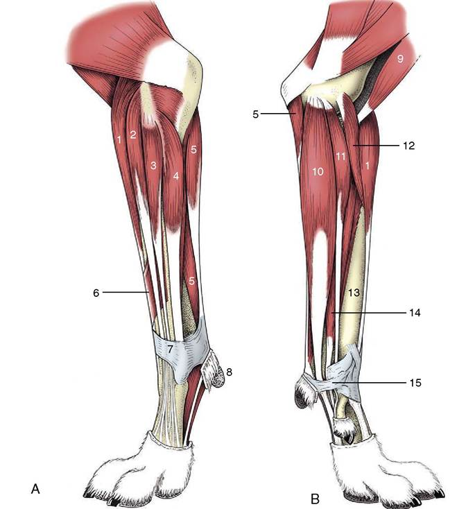
Figure 2-57 Muscles of the left forearm of the dog, lateral (A) and medial (B) views. 1, Extensor carpi radialis; 2, common digital extensor; 3, lateral digital extensor; 4, ulnaris lateralis; 5, flexor carpi ulnaris; 6, extensor carpi obliquus; 7, extensor retinaculum; 8, carpal pad; 9, biceps; 10, superficial digital flexor; 11, flexor carpi radialis; 12, pronator teres; 13, radius; 14, deep digital flexor; 15, flexor retinaculum.
into four in the pig and dog; and into five in the cat. A subdivision of the common extensor, which is present in all species but the horse and cat, inserts on the most medial of the functional digits; it sends an oblique branch to the dewclaw in the dog. It is sometimes usefully termed medial digital extensor, but this term is not official. The lateral digital extensor (Figure 2-57/3) runs along the lateral edge of the common extensor; the undivided tendon inserts on the dorsal surface of the proximal phalanx in the horse. The muscle also has one insertion tendon in the ruminants, two in the pig, three in the dog, and four in the cat; in these species the insertion is in common with the branch of the common extensor to the distal phalanx of the most lateral one, two, three, or four functional digits. In the smaller species, separation of the digital divisions begins more proximally and is more complete.
The Flexor Muscles of the Carpus and Digits. The carpal flexor group includes digital flexor muscles in addition to muscles that act only at the carpus. They have certain common features: a flexor action at the carpus; a caudal position in the forearm; an origin, in part at least, from the caudal aspect of the medial epicondyle of the humerus; and an innervation from the median or ulnar nerve, or from both these nerves. Some have additional, even principal, origins in the forearm and also act on the digital joints. The flexor carpi radialis (Figure 2-57/11) is most medial and runs directly caudal to the subcutaneous border of the radius. It ends on the upper end of the second (sometimes third) metacarpal bone. The flexor carpi ulnaris (Figure 2-57/5) is lateral and ends on the accessory carpal bone. Both muscles are solely carpal flexors.
The superficial digital flexor (Figure 2-57/10) lies in the caudomedial part of the forearm and is not enclosed in a synovial sheath where it passes the carpus; later it divides into a branch for each functional digit that inserts in the region of the proximal interphalangeal joint. To reach these positions the branches of the tendon must first change position with those of the deep flexor that continue to more distal terminations. In principle (although the details vary), each branch of the superficial flexor tendon splits into two slips that diverge to the sides of the deep tendon, which then passes through the resulting arch. The deep digital flexor (Figure 2-57/14) lies more deeply in the forearm and passes the carpus through the carpal canal before dividing into one to four digital branches; each perforates the corresponding branch of the superficial flexor tendon and then continues to its insertion on the palmar aspect of a distal phalanx.
Short Digital Muscles. Interosseous muscles support the metacarpophalangeal joints. They show marked species differences in number, structure (they are largely tendinous in the large species), and function. They arise from the palmar aspect of the proximal ends of the metacarpal bones and find initial insertion on the sesamoid bones at the metacarpophalangeal joints; from here they are continued by distal sesamoidean ligaments that attach to the phalanges and by extensor branches that wind around to the dorsal aspect of the digit to join the extensor tendons. They are considered in detail later for the species in which they are important.
In the carnivores and pig a number of small digital muscles assist in the extension, flexion, abduction, or adduction of the abaxial digits—one, two, and five in the dog and the cat and two and five in the pig. It is unnecessary to describe them.