THE JOINTS OF THE FORELIMB
The shoulder joint (Figure 2-52, A) links the scapula and humerus, and although it has attributes of the spheroidal variety, sagittal excursions predominate in practice. The glenoid cavity of the scapula is considerably smaller than the head of the humerus.
In large animals,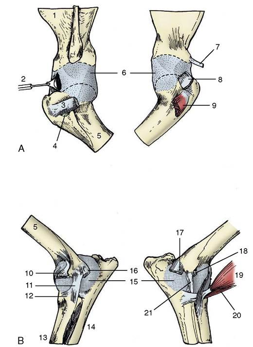
Figure 2-52 Left shoulder (A) and elbow (B) joints of the dog. The drawings on the left are lateral views, those on the right medial. 1, Scapula; 2, joint capsule opened to expose biceps tendon; 3, tendon of infraspinatus; 4, infraspinatus bursa; 5, humerus; 6, joint capsule, stretched by pulling bones apart; 7, tendon of coracobrachialis; 8, tendon of subscapularis, reflected ventrally; 9, biceps tendon emerging from intertubercular groove; 10, stump of extensor carpi radialis and common digital extensor; 11, lateral collateral ligament; 12, annular ligament of radius; 13, radius; 14, ulna; 15, joint capsule; 16, stump of ulnaris lateralis; 17, common stump of carpal and digital flexors; 18, stump of pronator teres; 19, biceps; 20, brachialis; 21, medial collateral ligament.
both surfaces may be indented peripherally by naked areas (synovial fossae) simulating, to the inexperienced eye, lesions of the cartilage. The joint capsule is roomy and is fused here and there with the tendons of the surrounding muscles, particularly the subscapularis. In all but the horse and ox it sends a prolongation or diverticulum around the tendon of origin of the biceps brachii, where this lies within the intertubercular groove. The diverticulum protects the tendon in the manner of a synovial sheath; it is replaced by a discrete intertubercular bursa in the two large species. Although the fibrous layer of the capsule is locally strengthened, it is usual to say that the joint is without pericapsular ligaments.
Tendons of immediately adjacent muscles, notably the subscapularis medially and infraspinatus laterally, take the place of ligaments in bracing the joint.Movement is most free in the sagittal direction, but significant amounts of rotation, abduction, and adduction, and therefore also of circumduction, are possible, particularly in the dog and cat; in these animals a component of the movement interpreted as supination probably occurs at shoulder level.
The elbow joint (Figure 2-52, B) combines within a single capsule the hinge joint between the humerus and the radius and ulna and, at least in carnivores, the pivot joint between the proximal extremities of the latter pair of bones. The humeral surface is broadly trochlear, and the lower surface, variously furnished by the radius and ulna, is its reciprocal. Ridging of the surfaces, most pronounced in the larger animals, impedes other than hinge movements. A proximal radioulnar articulation between a circumferential facet on the radius and a corresponding but smaller area on the ulna is present even when more distal fusion precludes the possibility of movement. The joint capsule is surprisingly roomy and, when distended, bulges to each side of the ulna within the olecranon fossa. The strongest ligaments are medial and lateral collateral ligaments, which is a predictable arrangement in what is basically a hinge joint.
The lateral of these ligaments is short and thick (Figure 2-52/11), and the medial one is longer, more slender, and divisible into two parts (Figure 2-52/21)— radial and ulnar in the dog and cat and superficial and deep in the larger animals. An additional oblique ligament is placed over the flexor aspect of the joint of the dog and cat. In these species there is also an annular ligament (Figure 2-52/12) extending between the collateral ligaments and completing the enclosure of the head of the radius within an osseoligamentous ring.
In the large species, most notably the horse, the curvature of the humeral surface is not uniform.
This feature, combined with the eccentric proximal attachment of the collateral ligaments (see Figure 23-10), makes the joint more stable in the normal standing position (which approaches but does not reach maximal extension); some effort is required to “unlock” the joint before it can be flexed.The shafts of the radius and ulna are joined by an interosseous membrane that ossifies early in life in ungulates. In the dog and cat the membrane is sufficiently long to allow the limited rotation possible in these species.
The carpal joint includes antebrachiocarpal, midcar- pal, and carpometacarpal levels of articulation and also a distal radioulnar joint. The antebrachiocarpal and the radioulnar joints share a common joint cavity. The mid- carpal and carpometacarpal joint cavities are interconnected. In hoofed species the proximal joint may be regarded as being of the hinge variety (although the form of the surfaces introduces a certain obliquity of movement in ruminants), but in dogs and cats it is more versatile and can be regarded as an ellipsoidal joint, although a poor example of the type. The hinge movement is quite free at the antebrachiocarpal level (horse: ca. 90°). Considerable movement is also possible at the midcarpal level (ca. 45°), but virtually no movement is allowed at the carpometacarpal level. Medial and lateral collateral ligaments are well developed in ungulates but are necessarily much weaker in the dog and cat to allow for some adduction and abduction. On the dorsal aspect, a number of short ligaments join neighboring bones in the same row and those of the row distal to the metacarpus. More robust ligaments are found on the palmar aspect, where a deep ligament (Figure 2—53/6) covers the entire palmar surface of the skeleton, burying the unevenness of the bones. A second, superficial, transverse ligament (flexor retinaculum) passes obliquely from the free extremity of the accessory carpal bone to the medial aspect of the carpus (Figure 2—53/7), completing the enclosure of a passage behind the carpus. This, the carpal canal, conveys the flexor tendons and other structures continuing into the foot from the forearm. Additional small ligaments (Figure 2—53/5) join the accessory bone to the adjacent carpal and metacarpal bones. These palmar ligaments do not interfere with flexion but assist in preventing overextension.
Description of the more distal joints is best deferred because of the marked interspecific variation. These joints are only important in the large species.