The Muscles of the Hindlimb
The girdle musculature has been described (p. 49).
The Intrinsic Muscles of the Hindlimb
Muscles Acting Primarily on the Hip Joint
The muscles acting at the hip are arranged topographically in gluteal, medial, deep, and caudal (hamstring) groups.
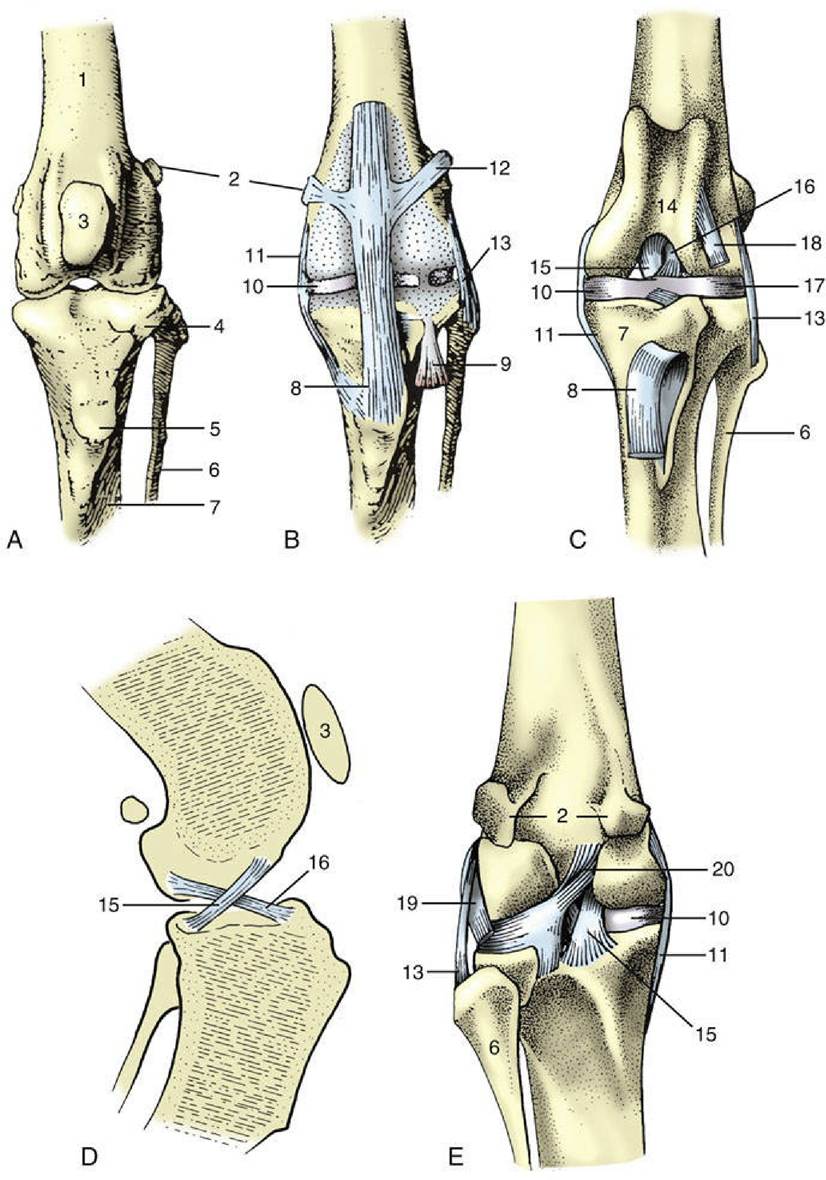
FIG.
2.63 (A to C) Left stifle joint of the dog, cranial views. (B) The extent of the joint capsule. (C) The patella has been removed. Crossing of the cruciate ligaments in (D) medial and (E) caudal views. 1, Femur; 2, sesamoids in the gastrocnemius; 3, patella; 4, extensor groove; 5, tibial tuberosity; 6, fibula; 7, tibia; 8, patellar ligament; 9, tendon of long digital extensor passing through extensor groove; 10, medial meniscus; 11, medial collateral ligament; 12, lateral femoropatellar ligament; 13, lateral collateral ligament; 14, trochlea; 15, caudal cruciate ligament; 16, cranial cruciate ligament; 17, lateral meniscus; 18, stump of 9; 19, popliteus tendon; 20, meniscofemoral ligament.The gluteal group comprises superficial, middle, and deep gluteal muscles and the tensor fasciae latae. The gluteus superficialis in the dog is a relatively narrow muscle that covers the caudal part
of the gluteus medius, extending from the gluteal and caudal fascia to the third trochanter of the femur (Fig. 2.64/4). In ungulates a part becomes incorporated within the biceps femoris, and sometimes also the semitendinosus, supplying them with vertebral heads of origin. The gluteus superficialis is an extensor of the hip and therefore a retractor of the limb. It is supplied by the caudal gluteal nerve.
The gluteus medius (Fig. 2.64/3) is by far the largest of the group. It arises from the outer surface of the ilium and the gluteal fascia and inserts on the greater trochanter. It is an exceptionally powerful extensor of the hip, with some abduction potential.
The actions of deeper subdivisions, the gluteus accessorius and the more caudal piriformis, are similar to those of the main mass. The muscle is principally supplied by the cranial gluteal nerve.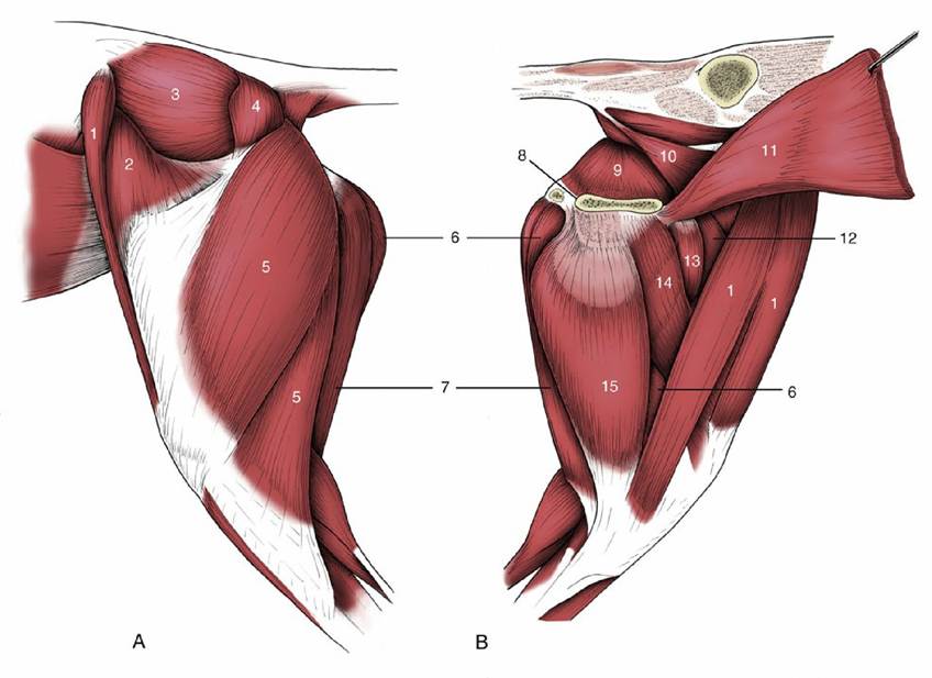
FIG. 2.64 Muscles of the canine hindquarter and thigh. (A) Lateral and (B) medial views. 1, Sartorius; 2, tensor fasciae latae; 3, gluteus medius; 4, gluteus superficialis; 5, biceps; 6, semimembranosus; 7, semitendinosus; 8, pelvic symphysis; 9, internal obturator; 10, levator ani; 11, rectus abdominis; 12, quadriceps; 13, pectineus; 14, adductor; 15, gracilis.
The much smaller gluteus profundus is completely covered by the gluteus medius. It arises from the ischial spine and adjacent region of the os coxae and inserts on the cranial part of the greater trochanter. It may also extend the hip, but the transverse course of most of its fibers makes it a more useful abductor. It is also supplied by the cranial gluteal nerve. The tensor fasciae latae (Fig. 2.64/2) is the most cranial muscle of the group. It arises from the coxal tuber (or equivalent) and from the adjacent part of the ilium and extends down the cranial border of the thigh before inserting into the heavy lateral femoral fascia, which provides it with attachment to the patella and other structures of the stifle region. Supplied by the cranial gluteal nerve, the tensor fasciae latae is primarily a flexor of the hip. In the horse its most caudal part extends toward and fuses with a cranial slip of the gluteus superficialis.
The medial group is principally employed to adduct the hindlimb; adduction is, of course, a term that also embraces the prevention of unwanted abduction. Most muscles of this group are supplied by the obturator nerve and they—gracilis, pectineus, adductor, and external obturator—are sometimes specifically termed the adductors. The sartorius has a rather different origin and relationship.
The gracilis, a broad but thin muscle, takes an aponeurotic origin from the symphyseal region of the pelvis (Fig.
2.64/15). Its insertion, also aponeurotic, merges with the crural fascia through which it finds attachment to the tibial crest and other medial structures of the stifle region.The pectineus is a small fusiform muscle, which in the dog forms a prominent surface feature of the proximal part of the thigh (Fig. 2.64/13). It arises from the cranial branch of the pubis and from the prepubic tendon and inserts on the proximal part of the medial "rough line" of the femur. In the larger species, but not in the dog, a considerable part of the tendon of origin decussates with its fellow within the prepubic tendon.
The adductor is often divided into several individually named parts, but these distinctions are unnecessary. The muscle arises over an extensive area of the ventral aspect of the pelvic floor and inserts along the distal two thirds of the medial "rough line" of the femur and to the fascia and ligaments of the medial aspect of the stifle (Fig. 2.64/14).
The obturator externus is conveniently included here, although it has obvious affinities with the deep group described next. It arises from the ventral surface of the pelvic floor, over and around the obturator foramen, and inserts within the ventral part of the trochanteric fossa. In addition to being an adductor, this muscle is potentially an outward rotator of the thigh.
The sartorius is set apart from the other medial muscles by its innervation from the saphenous branch of the femoral nerve. It is superficial and follows the craniomedial aspect of the thigh; in the dog it consists of two parallel bellies, one of which forms the cranial contour of the thigh (Fig. 2.64/1). Except in the horse (in which it arises from the iliac fascia on the abdominal roof), it arises from the iliac crest, and its insertion is to the medial structures of the stifle region. Flexion of the hip is probably its main action, but it has some capacity for adduction of the thigh and extension of the stifle. The superficial space between the caudal margin of the sartorius and the pectineus is often designated the femoral canal.
The deep muscles of the hip form a rather heterogeneous community of small and essentially trivial muscles: the obturator internus, gemelli, quadratus femoris, and articularis coxae. Most are supplied by the sciatic nerve.
The obturator internus (Fig. 2.64/9) is a thin muscle that arises from the dorsal surface of the femur in the vicinity of the obturator foramen; in carnivores and in the horse its tendon leaves the pelvis by passing over the ischium, caudal to the acetabulum, to end in the trochanteric fossa. In other species the tendon passes through the obturator foramen; in this arrangement, the muscle may have its origin as a detachment from the external obturator. The muscle is an external rotator of the thigh.
The gemelli are two small "twin" bundles that pass from the ischial spine to the trochanteric fossa. They are also external rotators.
The quadratus femoris passes from the ventral aspect of the ischium to end on the femoral shaft close to the trochanteric fossa. It is described as an extensor but can be of no significance in this role.
The articularis coxae lies on the capsule over the cranial aspect of the hip and protects it from being nipped between the femoral and acetabular surfaces.
The muscles of the caudal (hamstring) group—biceps femoris, semitendinosus, and semimembranosus—flesh the caudal part of the thigh. They extend from the ischial tuber and adjacent part of the sacrotuberous ligament to a broad insertion both proximal and distal to the joint space of the stifle; certain components continue within the common calcanean tendon to the calcaneus. In ungulates, one (or more) muscle(s) is also extended proximally through an origin (vertebral head) from the sacrocaudal vertebrae. The vertebral heads are best developed in the horse and account for the full, rounded contour of the rump of this animal, which contrasts with the more angular appearance in the ox or dog. At least part of the vertebral extension is due to assimilation of a superficial gluteal component.
The term gluteobiceps may be encountered for the combination.The biceps femoris is most lateral (Fig. 2.64/5). In the horse and in ruminants, but not in the dog, it has both vertebral and pelvic heads. In the lower part of the thigh the united muscle divides into insertions that attach, by way of the femoral and crural fascia, to the patella and ligaments of the stifle joint both proximal and distal to the joint space; an additional insertion to the point of the hock is achieved through a contribution (tarsal tendon) to the common calcanean tendon.
The semitendinosus (Fig. 2.64/7) forms the caudal contour of the thigh. It has a vertebral head only in the horse and pig. The insertion is to the medial aspect of the proximal extremity of the tibia and to the calcaneus. The insertions of the biceps and semitendinosus, one to each side of the depression (popliteal fossa) behind the stifle, can be palpated in life—they are the "strings of the ham" that give the group its name.
The semimembranosus (Fig. 2.64/6) is most medial and has a vertebral head only in the horse. The insertion is divided between a cranial part attaching to the medial femoral condyle and a caudal part attaching to the medial tibial condyle.
In the dog a ribbon-like and functionally insignificant abductor cruris caudalis lies on the deep face of the biceps and is probably derived from it.
The vertebral heads of these muscles are generally supplied by the caudal gluteal nerve, and the pelvic heads by the sciatic nerve (or its tibial division).
Collectively these muscles undoubtedly provide forceful extension of the hip joint that thrusts the trunk forward. In addition, the biceps has an abductor potential, and the semimembranosus an adductor potential, at the hip. When consideration is given to muscle action on the stifle, the points of insertion relative to the joint axis are more informative. it is probably more useful to divide the muscles into a cranial division inserting proximal to the joint axis and a caudal division inserting distal to this axis rather than to consider the named units.
Those cranial to the axis extend the stifle when the foot is planted on the ground, whereas the caudal division has the same action when the foot is fixed but flexes the joint when the foot is free to move. The parts of the biceps and semitendinosus that insert on the calcaneus can obviously extend the hock. All these effects, however, cannot be accomplished simultaneously because of both the potential antagonism of the cranial and caudal divisions at the stifle and the undesirability of flexing the stifle while extending the hock. Indeed, in the horse in particular, the reciprocal apparatus precludes this combination of actions (p. 625). Different parts of these muscles must therefore be used at different times and in different combinations.Muscles Acting Primarily on the Stifle Joint
There are extensor and flexor groups in the muscles acting on the stifle joint. The quadriceps femoris, the principal extensor of the stifle, forms the mass of muscle cranial to the femur (see Fig. 17.2/9). It originates as four parts but inserts as a single tendon distally. The rectus femoris originates from the shaft of the ilium immediately cranial to the acetabulum; the others, however—vastus medialis, intermedius, and lateralis—arise, respectively, from the medial, cranial, and lateral aspects of the femoral shaft. The common insertion appears to be on the patella but is actually on the tibial tuberosity because the muscle is continued distal to the patella by the patellar ligament(s). The rectus femoris has the potential secondary action of flexion of the hip. The quadriceps is supplied by the femoral nerve.
The small popliteus muscle lies directly over the caudal aspect of the joint. It takes a tendinous and confined origin from the lateral condyle of the femur and fans out to a broad fleshy insertion on the proximal third of the caudal surface of the tibia (Fig. 2.65/15). Its tendon of origin contains a sesamoid in the dog and cat. In addition to being a flexor of the stifle, the popliteus rotates the distal part of the limb. It is supplied by the tibial nerve.
Muscles Acting Primarily on the Tarsal and Digital Joints
The muscles acting on the tarsal and digital joints, which comprise extensors and flexors of the hock and the digits, are grouped in two masses: one craniolateral to the tibia and the other caudal to the tibia.
Craniolateral Muscles of the Leg
Some of the craniolateral group only flex the hock and others flex the hock and extend the digits. This arrangement contrasts with the digital extensors of the forelimb, which extend both carpal and distal joints. All of the craniolateral crural muscles are innervated through the peroneal* nerve (Fig. 2.65/3).
A full set of the muscles that are pure flexors of the hock are not found in any domestic species; it would consist of the tibialis cranialis, peroneus tertius, peroneus longus, and peroneus brevis.
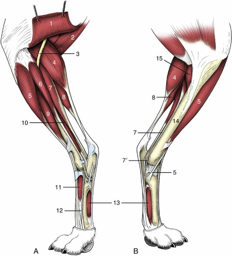
FIG. 2.65 Muscles of the left canine leg. (A) Lateral and (B) medial views. 1, Biceps; 2, semitendinosus; 3, peroneal nerve; 4, gastrocnemius; 5, tibialis cranialis; 6, peroneus longus; 7, lateral deep digital flexor; 7', tendon of the smaller medial deep digital flexor; 8, superficial digital flexor; 9, long digital extensor; 10, peroneus brevis; 11, extensor brevis; 12, tendon of lateral digital extensor; 13, interossei; 14, tibia; 15, popliteus.
The tibialis cranialis, always substantial, lies immediately cranial to the subcutaneous medial surface of the tibia (Fig. 2.65/5). After arising from the lateral condyle of the tibia, it inserts on the mediodistal tarsal and adjacent metatarsal skeleton. With hock flexion as its primary role, this muscle also is a supinator. The peroneus tertius is most important in the horse because of its critical role in the so-called reciprocal mechanism.
The weak peroneus longus arises from and around the distal part of the lateral collateral ligament of the stifle joint (Fig. 2.65/6). It crosses the lateral aspect of the tarsus before turning medially, over the plantar aspect, to end on the proximal parts of the medial metatarsal bone. It is primarily a pronator of the foot but may also flex the hock. The peroneus brevis is of no practical importance.
The number and the arrangement of the extensor muscles of the digits are naturally correlated with the digital pattern. A long digital extensor muscle (Fig. 2.65/9) arises from the distal extremity of the femur and follows the lateral border of the tibialis cranialis. Its tendon is held down by a retinaculum on the dorsal surface of the hock before it splits to send one branch for each functional digit. Each branch inserts on the extensor process of a distal phalanx. In the dog, the tendons develop small sesamoid bones similar to those in the forelimb.
A lateral digital extensor (Fig. 2.65/12) arises from the head of the fibula, crosses the lateral aspect of the hock, and enters the most lateral functional digit, where it terminates either on the proximal phalanx (dog) or by joining the long extensor tendon (horse). In certain species, including the dog, a small discrete extensor hallucis longus is associated with the medial digit; it arises on the cranial border of the fibula and inserts on the proximal part of the digit.
Caudal Muscles of the Leg
The caudal muscles of the leg are the twin-bellied gastrocnemius, the soleus, and the superficial and deep digital flexors. All are supplied by the tibial nerve.
The gastrocnemius and the soleus, the latter insignificant except in the cat and absent in the dog, are sometimes collectively known as the triceps surae. The two heads of the gastrocnemius (Fig. 2.65/4) spring from the caudal aspect of the femur proximal to the condyles; two sesamoid bones are included in their origins in carnivores. The heads combine in the upper part of the crus and give rise to a single stout tendon that inserts on the point of the hock. It is the principal component of the common calcanean (Achilles) tendon. Despite its inclusion among the extensors of the hock, the role of the gastrocnemius is enigmatic because its proximal attachment suggests that it is a potential flexor of the stifle; stifle and hock, however, normally move in unison. The apparent contradiction in these actions is not easily explained. It has been suggested that the prime function of the gastrocnemius is not to move either joint but to oppose bending of the tibia, ensuring that the strain is always directed along its long axis.
The superficial digital flexor (Fig. 2.65/8) arises from a supracondylar fossa or tubercle on the caudal aspect of the femur, close to the origin of the gastrocnemius. It first runs deeply, between the two parts of the latter muscle; its tendon later winds around the medial border of the gastrocnemius tendon to gain the more superficial position. It forms a broad cap over the point of the hock, where part finds attachment through medial and lateral slips, before continuing over the plantar aspect of the calcaneus to enter the foot; it is then disposed like the corresponding tendon of the forelimb. The muscle is heavily infiltrated by connective tissue, especially in the horse, in which it becomes almost entirely tendinous and forms the caudal component of the reciprocal mechanism.
There are three deep digital flexor muscles whose independence varies among species. The three —lateral and medial flexors and the tibialis caudalis—lie close together on the caudal surface of the tibia (and fibula), from which they take origin (Fig. 2.65/7). In the ungulates, the tendons of the lateral muscle and the tibialis caudalis unite above the tarsus and then run over the plantar aspect of the joint, medial to the calcaneus; this common tendon is then joined in the upper part of the metatarsus by that of the medial muscle, which descends over the medial malleolus. The combined deep flexor tendon ends as the corresponding tendon of the forelimb. In carnivores, only the lateral (Fig. 2.65/7) and medial (Fig. 2.65/7') muscles unite. The rather small tibialis caudalis remains aloof and inserts separately on the hock; this truncated course transforms it into an extensor of the hock and a supinator of the foot.
The most important short digital muscles are the interossei (Fig. 2.65/13), which resemble those of the forelimb. A number of other small muscles that occur, especially in the dog, are of trivial significance.
Comprehension Check
Develop a plan for integrated actions of bones and muscles of the limbs and the thorax that balance locomotion with respiration.
Develop a schematic view of musculoskeletal adaptations that facilitate quadrupeds to "carry" their heads.
What constitutes the carpal canal?
* Osteology derives from osteon, Greek (bone); arthrology from arthron, Greek (joint); and myology from mys, Greek (muscle). These terms, rather than the Latin equivalents, provide the stems for many medical terms: osteoma, arthrosis, myositis, and so forth. Syndesmology is sometimes used as an alternative term for the study of joints.
* This term is sometimes used elsewhere in a wider sense to include the mandible and even the hyoid apparatus. Because contemporary practice is inconsistent, a writer's intention must often be deduced from the context.
* The adjective fibular has equivalent meaning to peroneal and is substituted for it by some writers. At present, peroneal (in its Latin form, peroneus) is official.
158