The Joints of the Hindlimb
The hip joint (Fig. 2.62) is a spheroidal joint formed between the lunate surface of the acetabulum and the head of the femur. The acetabular surface is enlarged by an articular labrum (Fig.
2.62/2') continuous with the transverse acetabular ligament (Fig. 2.62/2'') that bridges the notch interrupting the medial wall of the socket. The synovial membrane of the joint is supported externally by a fibrous covering, which is not uniformly strong. The head of the femur is joined to the depth of the acetabulum by the intracapsular ligament of the femoral head, which is covered by a reflection of the synovial membrane. In the horse a second (accessory) ligament inserts on the nonarticular area of the head (p. 612).The hip does not enjoy the full range of movement expected of spheroidal joints. In the large animals movement is largely restricted to flexion and extension. In conformity with the dominance of sagittal movement, the articular area tends to extend onto the neck in ruminants. The restriction on movement is due to intraarticular ligament(s) and the massive medial muscles of the thigh. The joint has a more versatile range in the dog.
The stifle joint (Fig. 2.63), corresponding to the human knee, comprises femorotibial, femoropatellar, and proximal tibiofibular joints. The dog also has the joint between the femur and paired sesamoids in the origins of the gastrocnemius and the joint between the tibia and the sesamoid in the popliteus tendon. In the dog, all these articulations have a common synovial cavity, but in the large species the femoropatellar and the medial and lateral femorotibial compartments have more restricted communication with one another.
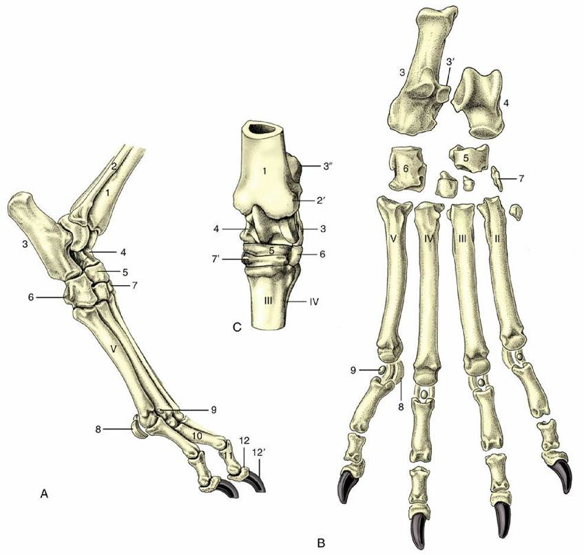
FIG. 2.61 Skeleton of right pes of the dog. (A) Lateral and (B) dorsal views. (C) Left equine tarsus, dorsal view.
Roman numerals identify the metatarsal bones. 1, Tibia; 2, fibula; 2', lateral malleolus; 3, calcaneus; 3', sustentaculum tali; 3", calcanean tuber (point of hock); 4, talus; 5, central tarsal; 6, fourth tarsal; 7, first, second, and third tarsal bones in distal row; 7', third tarsal in the horse; 8, proximal sesamoid bones; 9, dorsal sesamoid bones; 10, 11, and 12, proximal, middle, and distal phalanges; 12', claw.The femorotibial joint is unusual in having two fibrocartilaginous menisci (Fig. 2.63/10 and 17) interposed between the femoral and tibial condyles. The menisci, which compensate for the incongruence of the articular surfaces, are each semilunar in plan and wedge-shaped in section and have concave proximal and flattened distal surfaces. Each is secured by ligaments that extend between its cranial and caudal extremities and the central nonarticular area of the proximal extremity of the tibia; the lateral meniscus is also attached caudally to the intercondylar fossa of the femur.
Four ligaments join the femur to the bones of the leg. A medial collateral ligament passes between the femoral epicondyle and the proximal part of the tibia, toward the caudal part of the joint. The corresponding lateral ligament has a similar disposition but attaches to the fibular head. The cruciate ligaments are centrally placed. The cranial (lateral) cruciate ligament (Fig. 2.63/16) arises from the lateral condyle of the femur within the intercondylar fossa and runs craniodistally to attach on the tibia. The caudal (medial) cruciate ligament (Fig. 2.63/15) runs at right angles to the cranial one and attaches far back on the tibia near the popliteal notch.
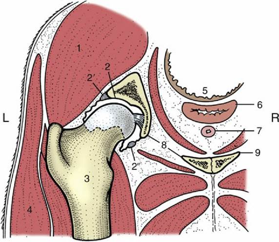
FIG. 2.62 Schematic transverse section through the left hip joint of a dog. The femur has been drawn in relief. 1, Gluteus medius; 2, acetabulum, connected to the femoral head by the ligament of the head of the femur; 2', fibrous rim (labrum) of acetabulum; 2", transverse acetabular ligament; 3, femur; 4, biceps; 5, rectum; 6, vagina; 7, urethra; 8, obturator foramen; 9, pelvic floor; L, left; R, right.
The femoropatellar joint is formed between the femoral trochlea and the patella and is extended by its parapatellar cartilages, the medial of which is especially well developed in the large animals.
Relatively weak collateral femoropatellar ligaments (Fig. 2.63/12) run between the cartilages and the femur. Distally the patella is joined to the tibial tuberosity by a single patellar ligament, except in the horse and ox, in which three ligamentous thickenings are present—medial, intermediate, and lateral-connected by a fibrous sheet (see Fig. 24.4). The middle (or single) patellar ligament represents the insertion tendon of the quadriceps femoris; the others, when present, represent the continuation of other muscles inserting about the joint.The synovial membrane attaches around the peripheries of the articular surfaces and the menisci. It covers the cruciate ligaments and here forms a partition, complete only in the horse, between the medial and lateral femorotibial joints. The femoropatellar portion of the cavity extends proximally between the femur and the quadriceps. In the horse it generally communicates only with the medial femorotibial compartment, but in other species it has free communication with both. Diverticula of the capsule embrace the lesser joints with the fibula and the sesamoid bones and extend along the tendons of origin of the long digital extensor and popliteus muscles.
Despite its complexity, the stifle functions as a hinge joint mainly entrusted with flexion and extension. The femoral condyles roll on the menisci, which in turn slide over the tibial plateau— cranially on extension, caudally on flexion. The travel between the femur and menisci is about three times that between the menisci and the tibia. The spiral configuration of the femoral condyles, when viewed from the side, tightens the ligaments and slows the movement when the joint moves toward the extended position. The stability of the articulation depends much on the cruciate ligaments. Rupture of one of these, which is not an uncommon misfortune, allows the tibia unusual mobility; it may slip forward when the cranial ligament is torn and backward when the caudal ligament is torn.
Rotation imposed on the joint, particularly when the joint is extended, places great strain on the menisci and their attachments.The tarsal joint of quadrupeds is usually known as the hock. Although it has four levels of articulation, in most species almost all movement occurs at the crurotarsal level. This is a hinge joint, but the obliquity of the interlocking ridges and grooves of the tibia and talus imposes a lateral deviation of the foot when it is carried forward on flexion. In ruminants and carnivores, limited flexion is also possible at the curved surfaces of the talocentral joint.
Among the many ligaments, the most important are the medial and lateral collateral ligaments, which extend, with intermediate attachments, from the tibia (and fibula) to the proximal extremity of the metatarsus. Each comprises a long superficial part of full extent and a shorter deeper part restricted to the proximal level of articulation. Another long ligament is found caudally, extending from the plantar surface of the calcaneus over the fourth tarsal bone to the metatarsus. The remaining smaller ligaments firmly hold the tarsal bones together.
There are several compartments to the joint. That between the tibia and talus is most capacious and may possess a number of local pouches, as the less supported parts of joint capsules are known. The other synovial sacs are much tighter and often communicate. The details are most important in the horse (p. 621).
The remaining joints of the hindlimb are considered in the regional chapters, insofar as they require to be differentiated from the corresponding forelimb joints.