The Nasal Cavity
The nasal cavity extends from the nostrils to the level of the eyes. Its rostral part, the nasal vestibule, is roughly tubular; caudal to the level of the infraorbital foramen, it widens and gains in height (Fig.
11.7). The nasal vestibule is occupied by the alar fold.The nasal cavity is divided into two halves by the nasal septum. In dogs, only the caudal and dorsal parts of the septum ossify; the rostral extremity projecting beyond the skull remains cartilaginous, accounting for the passive mobility of the tip of the nose. The middle section of the septum is membranous. A cat's nose is not actively mobile, and its cartilages resemble shortened canine nasal cartilages.
In dogs, the cavity is more tightly filled with nasal and ethmoidal conchae than in other species, and the intervening meatuses are narrow. The rostral half lodges the dorsal and ventral conchae. The dorsal one (Fig. 11.7/3) is a simple plate where it arises from the nasal bone, and it widens caudally to attach to the ethmoid. The ventral concha is thick but short, arises from the maxilla, and breaks into many scrolls that greatly enlarge the area that is covered by a richly vascularized mucosa (Fig. 11.7/2). The concha extends from the level of the first to the third premolar teeth and is attached to the conchal crest on the medial surface of the maxilla. This crest creates a linear shadow that is a very distinctive radiographic feature (Fig. 11.7B/14'). The ventral concha is continued rostrally by the alar fold. The caudal half of the nasal cavity is almost filled by ethmoidal conchae covered with olfactory mucosa. These conchae also invade the lower part of the frontal sinus. The olfactory mucosa in the German Shepherd reportedly covers an area of 150 cm2 and possesses more than 20 million receptors. The olfactory membrane differs little from the remainder of the mucous membrane, although it may be slightly thicker and grayer.
Collectively, the ethmoidal conchae are larger than the nasal conchae, a difference indicating the dog's keen sense of smell (see Fig. 11.10/11).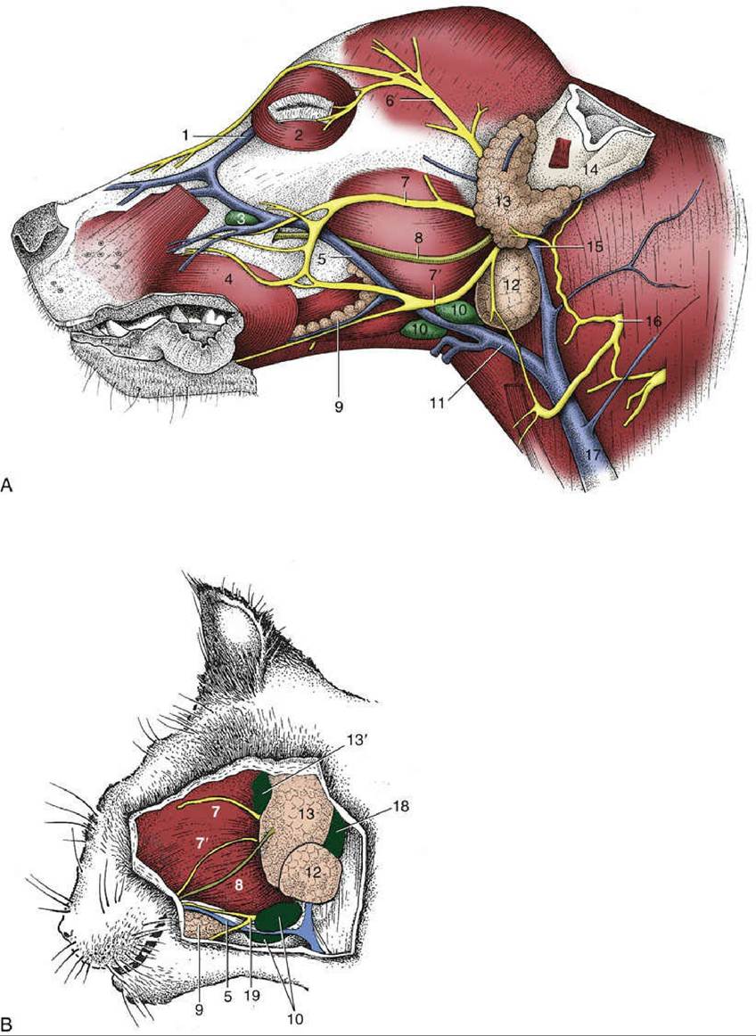
FIG. 11.6 Superficial dissection of (A) canine and (B) feline heads. 1, Angularis oculi vein; 2, orbicularis oculi; 3, facial lymph node; 4, orbicularis oris; 5, facial vein; 6, auriculopalpebral nerve; 7 and 7', dorsal and ventral buccal branches of facial nerve; 8, parotid duct; 9, buccal salivary glands; 10, mandibular lymph nodes; 11, linguofacial vein; 12, mandibular gland; 13, parotid gland; l3', parotid lymph node; 14, base of ear; 15, maxillary vein; 16, second cervical nerve; 17, external jugular vein; 18, lateral retropharyngeal lymph node; 19, facial nerve, ventral branch.
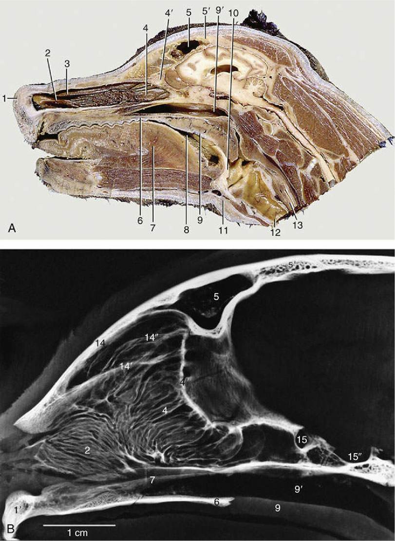
FIG. 11.7 (A) Paramedian section of the canine head. (B) Tomogram of the feline nasal cavity. 1, Right nostril; 2, ventral nasal concha; 3, dorsal nasal concha; 4, ethmoidal conchae; 4', cribriform plate; 5, frontal sinus; 5', frontal bone; 6, hard palate; 7, tongue; 7’, vomer; 8, oropharynx; 9, soft palate; 9', nasopharynx;
10, epiglottis; 11, basihyoid; 12, trachea; 13, esophagus; 14, nasal bone; 14', horizontal crest of nasal bone; 14", dorsal part of nasal cavity invaded by ethmoidal conchae; 15, optic canal; 15', hypophyseal fossa.
The nasal cavity of cats resembles the one of brachycephalic dogs. However, the ventral nasal concha is smaller, compensated for by enlargement and development of the middle concha and its lamellae. The middle concha reaches to the level of the entrance of the maxillary recess that it covers.
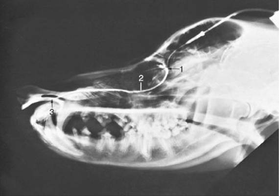
FIG. 11.8 Contrast medium outlining the canine nasolacrimal duct in a radiograph. 1, Position of ventral punctum; 2, nasolacrimal duct; 3, opening of duct at the nostril.
In both species, the nasolacrimal duct (Fig. 11.8) opens where the floor of the vestibule meets the alar fold and is visible when the nostril is spread. As often as not, there is a second, more caudal opening level with the canine tooth. The duct is described more extensively later. The duct of the lateral nasal gland opens at the rostral end of the dorsal nasal concha, but because it is only about 0.5 mm in diameter, it can be difficult to identify, even at dissection. The gland lies in the lateral nasal wall close to the entrance of the maxillary recess. Its secretion may have a social significance that accounts for the nose-to-nose sniffing common when dogs meet. In cats, the lateral nasal gland and its duct are not visible macroscopically; the secretion is mucous instead of serous.
A few much smaller nasal glands found on the rostral part of the septum open at the caudal limit of the vestibule and contribute marginally to the wetness of the nose. The watery secretions of the lacrimal, lateral nasal, and scattered minor nasal glands moisten the nasal plate.
The nasal cavity has an extremely good blood supply from both the external and internal carotid arteries; anastomoses occur between the internal carotid artery and the maxillary arteries (the main branch of the external carotid artery) of both sides. The maxillary artery is the major supply to the nasal cavity. Ligation of the external carotid artery in dogs (in cases of persistent nose bleeding) gives rise to collateral connections between corresponding vessels of both sides.