The neurological examination
To find out as much information as possible about a patient requires both keen observation before handling it, and then doing hands-on testing of neural functions. Begin with observation, then move to hands-on testing to assess function of the long tracts (proprioception, motor function), the cranial nerves, spinal reflexes, muscle tone and bulk and, finally, spinal hyperpathia.
This order of testing is from the most benign to the most noxious. A full physical examination must also be performed.Observation versus hands-on testing
The functioning of the majority of the nervous system can be assessed by close observation (Fig. 13.2). This is particularly useful if the animal cannot be handled; for example if it is fractious or wild. For animals that you can handle, then observation is still critical for getting an overall impression of neural function and is an excellent opportunity to assess arousal and behaviour. Physical contact usually stimulates the animal and subtle deficits in mental alertness may be missed. Assessing the animal’s arousal level is done best by observing its interaction with the environment. Answering the following questions provides useful information.
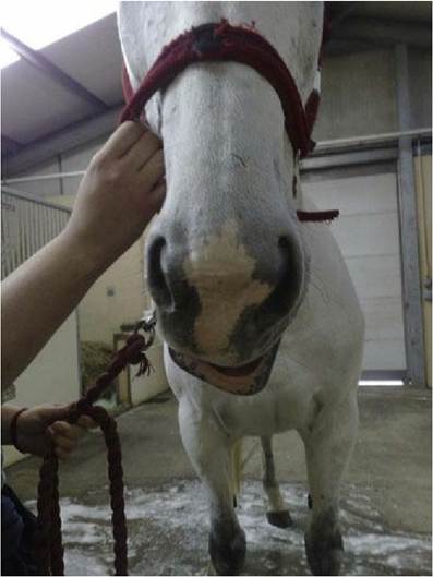
Fig. 13.2 What neurological deficit can be observed in this picture of a horse? (Answer on next page.)
Does it respond normally to environmental stimuli? These may include visual, auditory, olfactory, tactile and possibly gustatory stimuli. Does it seem bright, alert and responsive or dull and somnolent? (see Figs. 11.3 and 11.4)
Is the posture of its head, body, limbs and tail normal at rest and during locomotion? (See Fig. 6.2) Does it move normally or are there signs of proprioceptive deficits (stumbling, postural abnormalities), ataxia, stiffness, pain or paresis? If so, which limbs and which joints are functioning normally and which are dysfunctional? Is there any change in muscle bulk?
Observe the head closely.
Is the head posture normal? Is there any asymmetry of the face (drooping, muscle wasting)? (see Fig. 10.12) Do the ears, eyelids, eyes and nose move normally? Is it blinking? Is it sniffing at objects? Is it observing things or does it bump into them? Do both eyes track in a coordinated manner? Is there any strabismus, anisocoria or nystagmus? Does it respond to auditory stimuli? Is it swallowing normally or is there any evidence of respiratory stridor or change in voice? Is it licking its lips? Does it prehend food or drink normally?Are there any signs of autonomic dysfunction such as anisocoria, sweating, faecal or urinary incontinence? (see Fig. 12.6) Spinal reflexes are difficult to assess by observation, but the animal may show a skin twitch if an insect lands on it, or a perineal reflex after defecating or urinating. If the animal has normal posture and gait, then the spinal reflexes are likely to be normal.
The horse in Fig. 13.2 has a left-sided facial nerve paresis. This was due to accidental compression of the facial nerve by the head collar buckle while the horse was recumbent under anaesthesia (see Fig. 10.14B).
What do you observe in the horse in Fig. 13.3?
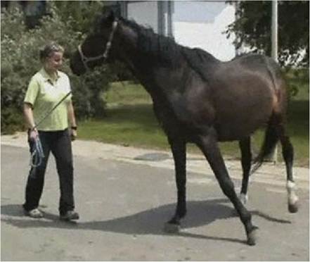
Fig. 13.3 What obvious type of deficit can be observed in this horse? (Answer in text.)
Proprioception and motor function
Normal gait requires both intact proprioception (limbs, trunk, head) and normal motor function (UMN and LMN). Similarly, for an animal to perform normally the proprioceptive tests used in the neurological examination, they require intact motor function. Therefore an animal with a purely motor problem could appear to have faulty proprioception, as it may not have the strength to place the limb in the correct position.
Assessing both proprioception and motor function is done by observing posture (trunk, limbs and head) and gait, and noting the positioning of the limbs under the centre of gravity, both at rest and during locomotion.
Are the limbs base-wide or base-narrow? Are the animal’s feet placed too far to the side or do they get crossed underneath while ambulating? Note also whether the animal bears weight on the correct part of the foot or has it knuckled over and is bearing weight on the dorsal aspect.If only the subconscious proprioception is compromised, then the limbs are often not placed under the centre of gravity either at rest or during locomotion. This results in base-wide or -narrow stance and ataxia with failure of the limbs to track under the centre of gravity when the animal is walking in a straight line. Deficits are often exaggerated when the animal turns. In horses this is seen particularly by excessive circumduction of the outside pelvic limb during tight turns.
The horse in Fig. 13.3 has proprioceptive deficits when turning in both the thoracic and the pelvic limbs. Note excessive circumduction of the left pelvic limb and the delayed movement of the left thoracic limb leaving the foot rotated inappropriately inwards.
If only conscious proprioception is compromised the animal may stand and walk with the limbs placed under the centre of gravity, but it may stand on top of the paw, or scuff the paw along the ground during the protraction phase, resulting in stumbling. Large animals, with their poorly developed corticospinal tracts, may have a remarkably normal gait with a forebrain injury, thus more complex manoeuvres are required to expose any deficit. What do you observe in the animals in Fig. 13.4?
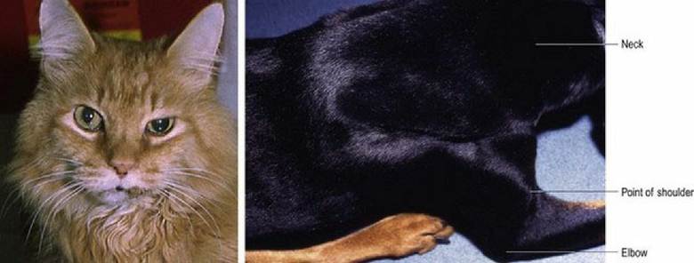
Fig. 13.4 What neurological deficits can be observed in these animals? (Answer in text.)
Cranial nerve function
Testing cranial nerve function by observation is outlined in Tables 10.2 and 13.1. Note that as cranial nerves are bilaterally paired, both sides of the head need to be checked (see Chapter 10).
Table 13.1 Observing cranial nerve function.
Note that blinking is a reflex action. The afferent stimulus is corneal drying stimulating CN V, ophthalmic branch to the brainstem and the efferent fibres travel in CN VII to the eyelids| Testing by observation | CNN being evaluated |
| Watch as the animal interacts with its environment | Many |
| Head position - tilted, the eyes are in a different plane compared with lateral rotation (torticollis) in which the eyes are in the same plane | Tilt - CN VIII (a), Vestibular |
| (Torticollis may be due to cervical or forebrain lesions) | |
| Facial symmetry, blinking, nostril and ear movement | CN VII |
| Blinking - due to stimulus of corneal drying | Ophthalmic branch of CN V (a), blinking due to CN VII (e) |
| Pupil size | Parasympathetic CN III (e) for miosis, or sympathetic (e) for mydriasis |
| Eyeball position | CNN II or VIII (a), CNN III, IV, VI (e) |
| Olfaction, vision, hearing, | CN I, II, VIII (a) |
| Masticatory muscle bulk, chewing | CN V (e) |
| Tongue movement, e.g. licking lips or nose | CN V (a) maxillary branch |
| CN XII (e) | |
| Swallowing | CN IX, X (a) and (e) |
| Laryngeal noise - phonation and stridor | CN X, XI (e) |
The cat has a dilated pupil in the left eye, subtle ventrolateral strabismus and loss of tone in the upper eyelid. This cat had CN III deficit causing loss of tone in the levator palpebrae superioris muscle (elevator of the upper eyelid), medial and dorsal rectus muscles (strabismus) and the iridal constrictor smooth muscle causing mydriasis.
This was due to a tumour in the floor of the cranial vault.The dog’s neurological deficits were readily detectable by observation. Clinically, the dog’s gait was ataxic (incoordinated) and paretic. It had marked atrophy of specific shoulder muscles, especially the supraspinatus muscle. In this particular case it was due to hypertrophied ligamentous tissue in the spinal canal compressing primarily the C6 spinal cord segment. The C6 spinal nerve arising from this segment is a key component of the suprascapular nerve innervating the supraspinatus muscle. LMNs supplying the suprascapular nerve and innervating the supraspinatus muscle (see Fig. 4.7). Functionally, loss of supraspinatus muscle function compromises shoulder extension resulting in hypometria (shortened strides) in the thoracic limbs. It also compressed sensory tracts and UMN tracts passing through the region supplying both the thoracic and pelvic limbs (see Fig. 4.8). Clinically the dog’s pelvic limb gait was ataxic (incoordinated - ‘wobbly’) and paretic, while the thoracic limb gait was short and stiff. This case was an example of rWobbler syndrome’ (see Fig. 6.3).